Youjin Jung, Kyu-Young Oh, Sang-Sun Han, Chena Lee
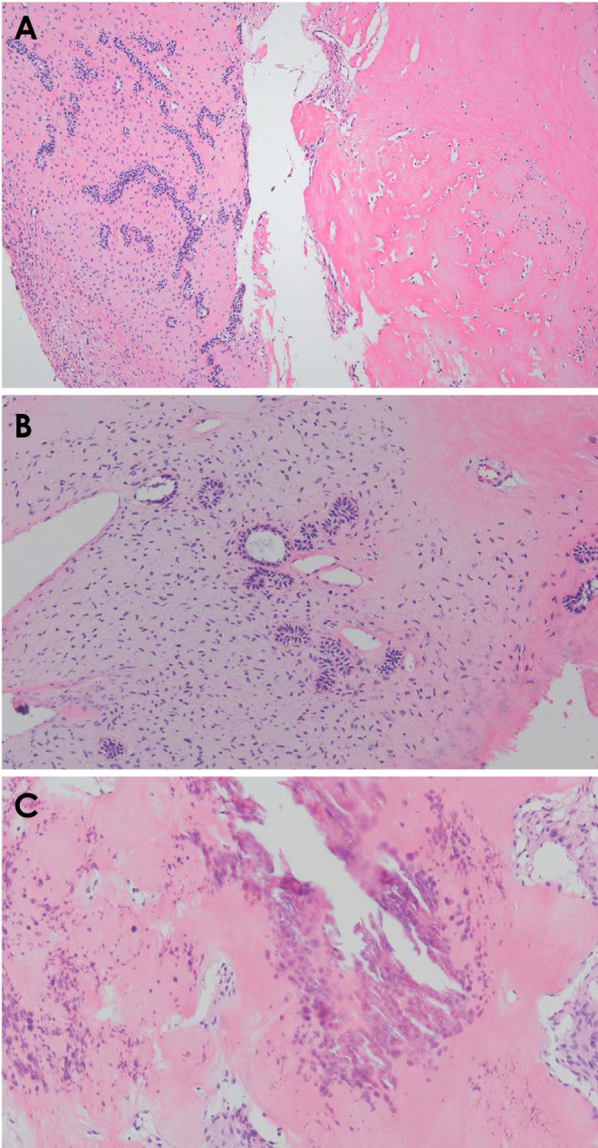
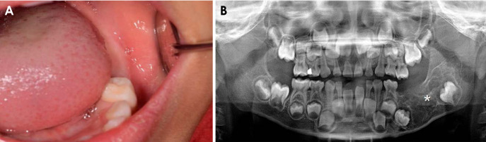
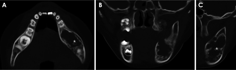
{"title":"A rare case report of ameloblastic fibrodentinoma with imaging features in a pediatric patient.","authors":"Youjin Jung, Kyu-Young Oh, Sang-Sun Han, Chena Lee","doi":"10.5624/isd.20230247","DOIUrl":null,"url":null,"abstract":"<p><p>Ameloblastic fibrodentinoma (AFD) is a rare benign odontogenic tumor that resembles an ameloblastic fibroma with dysplastic dentin. This report presents a rare case of mandibular AFD with imaging features in a young patient. Panoramic radiography and computed tomography revealed a well-defined lesion with internal septa and calcified foci, causing inferior displacement of the adjacent molars as well as buccolingual cortical thinning and expansion of the posterior mandible. The lesion was surgically removed via mass excision, and the involved tooth was extracted under general anesthesia. During the 5-year follow-up period, no evidence of recurrence was observed. Radiologic features of AFD typically reveal a moderately to well-defined mixed lesion with varying degrees of radiopacity, reflecting the extent of dentin formation. Radiologists should consider AFD in the differential diagnosis when encountering a multilocular lesion with little dense radiopacity, particularly if it is associated with delayed eruption, impaction, or absence of involved teeth, on radiographic images of young patients.</p>","PeriodicalId":51714,"journal":{"name":"Imaging Science in Dentistry","volume":"54 2","pages":"207-210"},"PeriodicalIF":2.1000,"publicationDate":"2024-06-01","publicationTypes":"Journal Article","fieldsOfStudy":null,"isOpenAccess":false,"openAccessPdf":"https://www.ncbi.nlm.nih.gov/pmc/articles/PMC11211030/pdf/","citationCount":"0","resultStr":null,"platform":"Semanticscholar","paperid":null,"PeriodicalName":"Imaging Science in Dentistry","FirstCategoryId":"1085","ListUrlMain":"https://doi.org/10.5624/isd.20230247","RegionNum":0,"RegionCategory":null,"ArticlePicture":[],"TitleCN":null,"AbstractTextCN":null,"PMCID":null,"EPubDate":"2024/4/2 0:00:00","PubModel":"Epub","JCR":"Q3","JCRName":"DENTISTRY, ORAL SURGERY & MEDICINE","Score":null,"Total":0}
引用次数: 0
Abstract
Ameloblastic fibrodentinoma (AFD) is a rare benign odontogenic tumor that resembles an ameloblastic fibroma with dysplastic dentin. This report presents a rare case of mandibular AFD with imaging features in a young patient. Panoramic radiography and computed tomography revealed a well-defined lesion with internal septa and calcified foci, causing inferior displacement of the adjacent molars as well as buccolingual cortical thinning and expansion of the posterior mandible. The lesion was surgically removed via mass excision, and the involved tooth was extracted under general anesthesia. During the 5-year follow-up period, no evidence of recurrence was observed. Radiologic features of AFD typically reveal a moderately to well-defined mixed lesion with varying degrees of radiopacity, reflecting the extent of dentin formation. Radiologists should consider AFD in the differential diagnosis when encountering a multilocular lesion with little dense radiopacity, particularly if it is associated with delayed eruption, impaction, or absence of involved teeth, on radiographic images of young patients.


 分享
分享
 求助内容:
求助内容: 应助结果提醒方式:
 扫码关注我们
