{"title":"Anterior talofibular ligament footprint dimension measured using three-dimensional magnetic resonance imaging.","authors":"Kenta Kono, Satoshi Yamaguchi, Seiji Kimura, Yukio Mikami, Kaoru Kitsukawa, Koji Matsumoto, Mutsuaki Edama, Yuki Shiko, Manato Horii, Takahisa Sasho, Seiji Ohtori","doi":"10.1007/s00256-024-04778-1","DOIUrl":null,"url":null,"abstract":"<p><strong>Objective: </strong>Knowledge of footprint anatomy is essential for ankle anterior talofibular ligament repair and reconstruction. We aimed to determine the intra- and inter-rater measurement reliability of the anterior talofibular ligament footprint dimension using three-dimensional MRI.</p><p><strong>Methods: </strong>MRI images of 20 ankles with intact ligaments, including 11 with a single bundle and nine with double-bundle ligaments, were analyzed. Imaging was performed using a 3.0-Tesla MRI. Isotropic three-dimensional proton density-weighted images with a voxel size of 0.6 mm were obtained. The fibular and talar footprints were manually segmented using image processing software to create three-dimensional ligament footprints. The lengths, widths, and areas of each sample were measured. A certified orthopedic surgeon and a senior orthopedic fellow performed the measurements twice at 6-week intervals. The intra- and inter-rater differences in the measurements were calculated.</p><p><strong>Results: </strong>The length, width, and area of the single-bundle fibular footprint were 8.7 mm, 5.4 mm, and 37.4 mm<sup>2</sup>, respectively. Those of the talar footprint were 8.4 mm, 4.3 mm, and 30.1 mm<sup>2</sup>, respectively. The inferior bundle of the double-bundle ligament was significantly smaller than the single and superior bundles (p < 0.001). No differences were observed between intra-rater measurements by either rater, with maximum differences of 0.7 mm, 0.5, and 1.7 mm<sup>2</sup>, in length, width, and area, respectively. The maximum inter-rater measurement differences were 1.9 mm, 0.5, and 2.4 mm<sup>2</sup>, respectively.</p><p><strong>Conclusion: </strong>Measurements of the anterior talofibular ligament dimensions using three-dimensional MRI were sufficiently reliable. This measurement method provides in vivo quantitative data on ligament footprint anatomy.</p>","PeriodicalId":21783,"journal":{"name":"Skeletal Radiology","volume":null,"pages":null},"PeriodicalIF":1.9000,"publicationDate":"2024-09-07","publicationTypes":"Journal Article","fieldsOfStudy":null,"isOpenAccess":false,"openAccessPdf":"","citationCount":"0","resultStr":null,"platform":"Semanticscholar","paperid":null,"PeriodicalName":"Skeletal Radiology","FirstCategoryId":"3","ListUrlMain":"https://doi.org/10.1007/s00256-024-04778-1","RegionNum":3,"RegionCategory":"医学","ArticlePicture":[],"TitleCN":null,"AbstractTextCN":null,"PMCID":null,"EPubDate":"","PubModel":"","JCR":"Q2","JCRName":"ORTHOPEDICS","Score":null,"Total":0}
引用次数: 0
Abstract
Objective: Knowledge of footprint anatomy is essential for ankle anterior talofibular ligament repair and reconstruction. We aimed to determine the intra- and inter-rater measurement reliability of the anterior talofibular ligament footprint dimension using three-dimensional MRI.
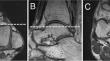
Methods: MRI images of 20 ankles with intact ligaments, including 11 with a single bundle and nine with double-bundle ligaments, were analyzed. Imaging was performed using a 3.0-Tesla MRI. Isotropic three-dimensional proton density-weighted images with a voxel size of 0.6 mm were obtained. The fibular and talar footprints were manually segmented using image processing software to create three-dimensional ligament footprints. The lengths, widths, and areas of each sample were measured. A certified orthopedic surgeon and a senior orthopedic fellow performed the measurements twice at 6-week intervals. The intra- and inter-rater differences in the measurements were calculated.
Results: The length, width, and area of the single-bundle fibular footprint were 8.7 mm, 5.4 mm, and 37.4 mm2, respectively. Those of the talar footprint were 8.4 mm, 4.3 mm, and 30.1 mm2, respectively. The inferior bundle of the double-bundle ligament was significantly smaller than the single and superior bundles (p < 0.001). No differences were observed between intra-rater measurements by either rater, with maximum differences of 0.7 mm, 0.5, and 1.7 mm2, in length, width, and area, respectively. The maximum inter-rater measurement differences were 1.9 mm, 0.5, and 2.4 mm2, respectively.
Conclusion: Measurements of the anterior talofibular ligament dimensions using three-dimensional MRI were sufficiently reliable. This measurement method provides in vivo quantitative data on ligament footprint anatomy.
期刊介绍:
Skeletal Radiology provides a forum for the dissemination of current knowledge and information dealing with disorders of the musculoskeletal system including the spine. While emphasizing the radiological aspects of the many varied skeletal abnormalities, the journal also adopts an interdisciplinary approach, reflecting the membership of the International Skeletal Society. Thus, the anatomical, pathological, physiological, clinical, metabolic and epidemiological aspects of the many entities affecting the skeleton receive appropriate consideration.
This is the Journal of the International Skeletal Society and the Official Journal of the Society of Skeletal Radiology and the Australasian Musculoskelelal Imaging Group.


 分享
分享
 求助内容:
求助内容: 应助结果提醒方式:
 扫码关注我们
