Romario Gorgis, Søren Aksel Christian Krarup, Jesper Reibel, Sven Erik Nørholt
{"title":"腺源性牙源性囊肿1例报告并文献复习。","authors":"Romario Gorgis, Søren Aksel Christian Krarup, Jesper Reibel, Sven Erik Nørholt","doi":"10.5037/jomr.2023.14204","DOIUrl":null,"url":null,"abstract":"<p><strong>Background: </strong>The glandular odontogenic cyst is now a well-known entity comprising < 0.5% of all odontogenic cysts with a recent review tabulating about 200 cases in the English literature. Glandular odontogenic cyst shows epithelial features that simulate salivary gland or glandular differentiation. The importance of glandular odontogenic cyst relates to the fact that it has a high recurrence rate and shares overlapping histologic features with central mucoepidermoid carcinoma. The purpose of this paper is to describe the clinical, radiological, and histopathological features of a case of glandular odontogenic cyst with the course of treatment and 9-years follow-up, followed by a review of the literature.</p><p><strong>Methods: </strong>A 63-year-old male was referred for further investigation of a mandibular radiolucency observed by his general dental practitioner. The main complaint was a murmuring sensation in the lower jaw right side. Radiological examination revealed a well-defined, unilocular, radiolucent lesion, involving the right mandible with 17 and 68 mm in mediolaterally and anteroposterior dimension, respectively.</p><p><strong>Results: </strong>A total enucleation of the cystic lesion and surgical extraction of tooth #46, #47 and #48, was performed under local anaesthesia. Histopathologic examination revealed a glandular odontogenic cyst.</p><p><strong>Conclusions: </strong>Glandular odontogenic cyst shows no pathognomonic clinico-radiographic characteristics, and therefore in many cases it resembles a wide spectrum of lesions. Diagnosis can be extremely difficult due to histopathological similarities with dentigerous cyst, lateral periodontal cyst and central mucoepidermoid carcinoma. Therefore a careful histopathological examination and a long-term follow-up (preferably seven years) are required to rule out recurrences.</p>","PeriodicalId":53254,"journal":{"name":"eJournal of Oral Maxillofacial Research","volume":null,"pages":null},"PeriodicalIF":1.0000,"publicationDate":"2023-04-01","publicationTypes":"Journal Article","fieldsOfStudy":null,"isOpenAccess":false,"openAccessPdf":"https://ftp.ncbi.nlm.nih.gov/pub/pmc/oa_pdf/82/46/jomr-14-e4.PMC10382194.pdf","citationCount":"0","resultStr":"{\"title\":\"Glandular Odontogenic Cyst: a Case Report and Literature Review.\",\"authors\":\"Romario Gorgis, Søren Aksel Christian Krarup, Jesper Reibel, Sven Erik Nørholt\",\"doi\":\"10.5037/jomr.2023.14204\",\"DOIUrl\":null,\"url\":null,\"abstract\":\"<p><strong>Background: </strong>The glandular odontogenic cyst is now a well-known entity comprising < 0.5% of all odontogenic cysts with a recent review tabulating about 200 cases in the English literature. Glandular odontogenic cyst shows epithelial features that simulate salivary gland or glandular differentiation. The importance of glandular odontogenic cyst relates to the fact that it has a high recurrence rate and shares overlapping histologic features with central mucoepidermoid carcinoma. The purpose of this paper is to describe the clinical, radiological, and histopathological features of a case of glandular odontogenic cyst with the course of treatment and 9-years follow-up, followed by a review of the literature.</p><p><strong>Methods: </strong>A 63-year-old male was referred for further investigation of a mandibular radiolucency observed by his general dental practitioner. The main complaint was a murmuring sensation in the lower jaw right side. Radiological examination revealed a well-defined, unilocular, radiolucent lesion, involving the right mandible with 17 and 68 mm in mediolaterally and anteroposterior dimension, respectively.</p><p><strong>Results: </strong>A total enucleation of the cystic lesion and surgical extraction of tooth #46, #47 and #48, was performed under local anaesthesia. Histopathologic examination revealed a glandular odontogenic cyst.</p><p><strong>Conclusions: </strong>Glandular odontogenic cyst shows no pathognomonic clinico-radiographic characteristics, and therefore in many cases it resembles a wide spectrum of lesions. Diagnosis can be extremely difficult due to histopathological similarities with dentigerous cyst, lateral periodontal cyst and central mucoepidermoid carcinoma. Therefore a careful histopathological examination and a long-term follow-up (preferably seven years) are required to rule out recurrences.</p>\",\"PeriodicalId\":53254,\"journal\":{\"name\":\"eJournal of Oral Maxillofacial Research\",\"volume\":null,\"pages\":null},\"PeriodicalIF\":1.0000,\"publicationDate\":\"2023-04-01\",\"publicationTypes\":\"Journal Article\",\"fieldsOfStudy\":null,\"isOpenAccess\":false,\"openAccessPdf\":\"https://ftp.ncbi.nlm.nih.gov/pub/pmc/oa_pdf/82/46/jomr-14-e4.PMC10382194.pdf\",\"citationCount\":\"0\",\"resultStr\":null,\"platform\":\"Semanticscholar\",\"paperid\":null,\"PeriodicalName\":\"eJournal of Oral Maxillofacial Research\",\"FirstCategoryId\":\"1085\",\"ListUrlMain\":\"https://doi.org/10.5037/jomr.2023.14204\",\"RegionNum\":0,\"RegionCategory\":null,\"ArticlePicture\":[],\"TitleCN\":null,\"AbstractTextCN\":null,\"PMCID\":null,\"EPubDate\":\"\",\"PubModel\":\"\",\"JCR\":\"Q3\",\"JCRName\":\"DENTISTRY, ORAL SURGERY & MEDICINE\",\"Score\":null,\"Total\":0}","platform":"Semanticscholar","paperid":null,"PeriodicalName":"eJournal of Oral Maxillofacial Research","FirstCategoryId":"1085","ListUrlMain":"https://doi.org/10.5037/jomr.2023.14204","RegionNum":0,"RegionCategory":null,"ArticlePicture":[],"TitleCN":null,"AbstractTextCN":null,"PMCID":null,"EPubDate":"","PubModel":"","JCR":"Q3","JCRName":"DENTISTRY, ORAL SURGERY & MEDICINE","Score":null,"Total":0}
Glandular Odontogenic Cyst: a Case Report and Literature Review.
Background: The glandular odontogenic cyst is now a well-known entity comprising < 0.5% of all odontogenic cysts with a recent review tabulating about 200 cases in the English literature. Glandular odontogenic cyst shows epithelial features that simulate salivary gland or glandular differentiation. The importance of glandular odontogenic cyst relates to the fact that it has a high recurrence rate and shares overlapping histologic features with central mucoepidermoid carcinoma. The purpose of this paper is to describe the clinical, radiological, and histopathological features of a case of glandular odontogenic cyst with the course of treatment and 9-years follow-up, followed by a review of the literature.
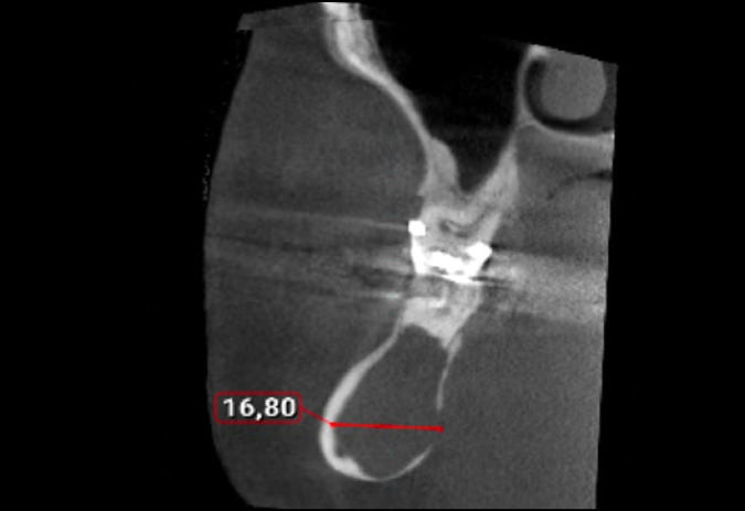
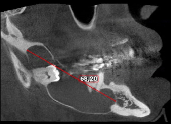
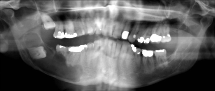
Methods: A 63-year-old male was referred for further investigation of a mandibular radiolucency observed by his general dental practitioner. The main complaint was a murmuring sensation in the lower jaw right side. Radiological examination revealed a well-defined, unilocular, radiolucent lesion, involving the right mandible with 17 and 68 mm in mediolaterally and anteroposterior dimension, respectively.
Results: A total enucleation of the cystic lesion and surgical extraction of tooth #46, #47 and #48, was performed under local anaesthesia. Histopathologic examination revealed a glandular odontogenic cyst.
Conclusions: Glandular odontogenic cyst shows no pathognomonic clinico-radiographic characteristics, and therefore in many cases it resembles a wide spectrum of lesions. Diagnosis can be extremely difficult due to histopathological similarities with dentigerous cyst, lateral periodontal cyst and central mucoepidermoid carcinoma. Therefore a careful histopathological examination and a long-term follow-up (preferably seven years) are required to rule out recurrences.


 分享
分享

 求助内容:
求助内容: 应助结果提醒方式:
 扫码关注我们
