Janna E. G. Roet, Aleksandra M. Mikula, Michael de Kok, Cora H. Chadick, Juan J. Garcia Vallejo, Henk P. Roest, Luc J. W. van der Laan, Charlotte M. de Winde, Reina E. Mebius
{"title":"对具有多种自发荧光光谱的细胞进行光谱分析的无偏方法。","authors":"Janna E. G. Roet, Aleksandra M. Mikula, Michael de Kok, Cora H. Chadick, Juan J. Garcia Vallejo, Henk P. Roest, Luc J. W. van der Laan, Charlotte M. de Winde, Reina E. Mebius","doi":"10.1002/cyto.a.24856","DOIUrl":null,"url":null,"abstract":"<p>Autofluorescence is an intrinsic feature of cells, caused by the natural emission of light by photo-excitatory molecular content, which can complicate analysis of flow cytometry data. Different cell types have different autofluorescence spectra and, even within one cell type, heterogeneity of autofluorescence spectra can be present, for example, as a consequence of activation status or metabolic changes. By using full spectrum flow cytometry, the emission spectrum of a fluorochrome is captured by a set of photo detectors across a range of wavelengths, creating an unique signature for that fluorochrome. This signature is then used to identify, or unmix, that fluorochrome's unique spectrum from a multicolor sample containing different fluorescent molecules. Importantly, this means that this technology can also be used to identify intrinsic autofluorescence signal of an unstained sample, which can be used for unmixing purposes and to separate the autofluorescence signal from the fluorophore signals. However, this only works if the sample has a singular, relatively homogeneous and bright autofluorescence spectrum. To analyze samples with heterogeneous autofluorescence spectral profiles, we setup an unbiased workflow to more quickly identify differing autofluorescence spectra present in a sample to include as “autofluorescence signatures” during the unmixing of the full stained samples. First, clusters of cells with similar autofluorescence spectra are identified by unbiased dimensional reduction and clustering of unstained cells. Then, unique autofluorescence clusters are determined and are used to improve the unmixing accuracy of the full stained sample. Independent of the intensity of the autofluorescence and immunophenotyping of cell subsets, this unbiased method allows for the identification of most of the distinct autofluorescence spectra present in a sample, leading to less confounding autofluorescence spillover and spread into extrinsic phenotyping markers. Furthermore, this method is equally useful for spectral analysis of different biological samples, including tissue cell suspensions, peripheral blood mononuclear cells, and in vitro cultures of (primary) cells.</p>","PeriodicalId":11068,"journal":{"name":"Cytometry Part A","volume":null,"pages":null},"PeriodicalIF":2.5000,"publicationDate":"2024-06-12","publicationTypes":"Journal Article","fieldsOfStudy":null,"isOpenAccess":false,"openAccessPdf":"https://onlinelibrary.wiley.com/doi/epdf/10.1002/cyto.a.24856","citationCount":"0","resultStr":"{\"title\":\"Unbiased method for spectral analysis of cells with great diversity of autofluorescence spectra\",\"authors\":\"Janna E. G. Roet, Aleksandra M. Mikula, Michael de Kok, Cora H. Chadick, Juan J. Garcia Vallejo, Henk P. Roest, Luc J. W. van der Laan, Charlotte M. de Winde, Reina E. Mebius\",\"doi\":\"10.1002/cyto.a.24856\",\"DOIUrl\":null,\"url\":null,\"abstract\":\"<p>Autofluorescence is an intrinsic feature of cells, caused by the natural emission of light by photo-excitatory molecular content, which can complicate analysis of flow cytometry data. Different cell types have different autofluorescence spectra and, even within one cell type, heterogeneity of autofluorescence spectra can be present, for example, as a consequence of activation status or metabolic changes. By using full spectrum flow cytometry, the emission spectrum of a fluorochrome is captured by a set of photo detectors across a range of wavelengths, creating an unique signature for that fluorochrome. This signature is then used to identify, or unmix, that fluorochrome's unique spectrum from a multicolor sample containing different fluorescent molecules. Importantly, this means that this technology can also be used to identify intrinsic autofluorescence signal of an unstained sample, which can be used for unmixing purposes and to separate the autofluorescence signal from the fluorophore signals. However, this only works if the sample has a singular, relatively homogeneous and bright autofluorescence spectrum. To analyze samples with heterogeneous autofluorescence spectral profiles, we setup an unbiased workflow to more quickly identify differing autofluorescence spectra present in a sample to include as “autofluorescence signatures” during the unmixing of the full stained samples. First, clusters of cells with similar autofluorescence spectra are identified by unbiased dimensional reduction and clustering of unstained cells. Then, unique autofluorescence clusters are determined and are used to improve the unmixing accuracy of the full stained sample. Independent of the intensity of the autofluorescence and immunophenotyping of cell subsets, this unbiased method allows for the identification of most of the distinct autofluorescence spectra present in a sample, leading to less confounding autofluorescence spillover and spread into extrinsic phenotyping markers. Furthermore, this method is equally useful for spectral analysis of different biological samples, including tissue cell suspensions, peripheral blood mononuclear cells, and in vitro cultures of (primary) cells.</p>\",\"PeriodicalId\":11068,\"journal\":{\"name\":\"Cytometry Part A\",\"volume\":null,\"pages\":null},\"PeriodicalIF\":2.5000,\"publicationDate\":\"2024-06-12\",\"publicationTypes\":\"Journal Article\",\"fieldsOfStudy\":null,\"isOpenAccess\":false,\"openAccessPdf\":\"https://onlinelibrary.wiley.com/doi/epdf/10.1002/cyto.a.24856\",\"citationCount\":\"0\",\"resultStr\":null,\"platform\":\"Semanticscholar\",\"paperid\":null,\"PeriodicalName\":\"Cytometry Part A\",\"FirstCategoryId\":\"99\",\"ListUrlMain\":\"https://onlinelibrary.wiley.com/doi/10.1002/cyto.a.24856\",\"RegionNum\":4,\"RegionCategory\":\"生物学\",\"ArticlePicture\":[],\"TitleCN\":null,\"AbstractTextCN\":null,\"PMCID\":null,\"EPubDate\":\"\",\"PubModel\":\"\",\"JCR\":\"Q3\",\"JCRName\":\"BIOCHEMICAL RESEARCH METHODS\",\"Score\":null,\"Total\":0}","platform":"Semanticscholar","paperid":null,"PeriodicalName":"Cytometry Part A","FirstCategoryId":"99","ListUrlMain":"https://onlinelibrary.wiley.com/doi/10.1002/cyto.a.24856","RegionNum":4,"RegionCategory":"生物学","ArticlePicture":[],"TitleCN":null,"AbstractTextCN":null,"PMCID":null,"EPubDate":"","PubModel":"","JCR":"Q3","JCRName":"BIOCHEMICAL RESEARCH METHODS","Score":null,"Total":0}
Unbiased method for spectral analysis of cells with great diversity of autofluorescence spectra
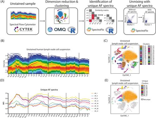
Autofluorescence is an intrinsic feature of cells, caused by the natural emission of light by photo-excitatory molecular content, which can complicate analysis of flow cytometry data. Different cell types have different autofluorescence spectra and, even within one cell type, heterogeneity of autofluorescence spectra can be present, for example, as a consequence of activation status or metabolic changes. By using full spectrum flow cytometry, the emission spectrum of a fluorochrome is captured by a set of photo detectors across a range of wavelengths, creating an unique signature for that fluorochrome. This signature is then used to identify, or unmix, that fluorochrome's unique spectrum from a multicolor sample containing different fluorescent molecules. Importantly, this means that this technology can also be used to identify intrinsic autofluorescence signal of an unstained sample, which can be used for unmixing purposes and to separate the autofluorescence signal from the fluorophore signals. However, this only works if the sample has a singular, relatively homogeneous and bright autofluorescence spectrum. To analyze samples with heterogeneous autofluorescence spectral profiles, we setup an unbiased workflow to more quickly identify differing autofluorescence spectra present in a sample to include as “autofluorescence signatures” during the unmixing of the full stained samples. First, clusters of cells with similar autofluorescence spectra are identified by unbiased dimensional reduction and clustering of unstained cells. Then, unique autofluorescence clusters are determined and are used to improve the unmixing accuracy of the full stained sample. Independent of the intensity of the autofluorescence and immunophenotyping of cell subsets, this unbiased method allows for the identification of most of the distinct autofluorescence spectra present in a sample, leading to less confounding autofluorescence spillover and spread into extrinsic phenotyping markers. Furthermore, this method is equally useful for spectral analysis of different biological samples, including tissue cell suspensions, peripheral blood mononuclear cells, and in vitro cultures of (primary) cells.
期刊介绍:
Cytometry Part A, the journal of quantitative single-cell analysis, features original research reports and reviews of innovative scientific studies employing quantitative single-cell measurement, separation, manipulation, and modeling techniques, as well as original articles on mechanisms of molecular and cellular functions obtained by cytometry techniques.
The journal welcomes submissions from multiple research fields that fully embrace the study of the cytome:
Biomedical Instrumentation Engineering
Biophotonics
Bioinformatics
Cell Biology
Computational Biology
Data Science
Immunology
Parasitology
Microbiology
Neuroscience
Cancer
Stem Cells
Tissue Regeneration.


 分享
分享
 求助内容:
求助内容: 应助结果提醒方式:
 扫码关注我们
