{"title":"胶质母细胞瘤,idh野生型伴脑膜轻脑膜转移至Meckel穴1例。","authors":"Toshiki Murata, Masazumi Matsuda, Tetsugaku Shinozaki, Koichi Ishiyama","doi":"10.1177/20584601221131480","DOIUrl":null,"url":null,"abstract":"<p><p>Meckel's cave or the trigeminal cistern is a subarachnoid space near the apex of the petrous portion of the temporal bone and contains cerebrospinal fluid and the Gasserian ganglion, which divides into the ophthalmic (V1), maxillary (V2), and mandibular (V3) nerves. Infectious, inflammatory, congenital, and neoplastic lesions can occur in Meckel's cave. Leptomeningeal metastasis of glioblastoma (GBM), IDH-wildtype to Meckel's cave is rare. We encountered a case of leptomeningeal metastasis of GBM to Meckel's cave in an elderly female patient who presented with pain around her right eye. Magnetic resonance imaging revealed enhancing lesions in the right temporal lobe and cervical spinal cord. The pathological diagnosis of GBM was confirmed after biopsy of the cervical spinal cord lesion, which showed hyperaccumulation of fluorodeoxyglucose (FDG) on FDG-positron emission tomography. This case indicates that metastatic lesions can also occur in Meckel's cave.</p>","PeriodicalId":72063,"journal":{"name":"Acta radiologica open","volume":null,"pages":null},"PeriodicalIF":0.9000,"publicationDate":"2022-10-07","publicationTypes":"Journal Article","fieldsOfStudy":null,"isOpenAccess":false,"openAccessPdf":"https://ftp.ncbi.nlm.nih.gov/pub/pmc/oa_pdf/72/bc/10.1177_20584601221131480.PMC9549091.pdf","citationCount":"1","resultStr":"{\"title\":\"Glioblastoma, IDH-wildtype with leptomeningeal metastasis to Meckel's cave: A case report.\",\"authors\":\"Toshiki Murata, Masazumi Matsuda, Tetsugaku Shinozaki, Koichi Ishiyama\",\"doi\":\"10.1177/20584601221131480\",\"DOIUrl\":null,\"url\":null,\"abstract\":\"<p><p>Meckel's cave or the trigeminal cistern is a subarachnoid space near the apex of the petrous portion of the temporal bone and contains cerebrospinal fluid and the Gasserian ganglion, which divides into the ophthalmic (V1), maxillary (V2), and mandibular (V3) nerves. Infectious, inflammatory, congenital, and neoplastic lesions can occur in Meckel's cave. Leptomeningeal metastasis of glioblastoma (GBM), IDH-wildtype to Meckel's cave is rare. We encountered a case of leptomeningeal metastasis of GBM to Meckel's cave in an elderly female patient who presented with pain around her right eye. Magnetic resonance imaging revealed enhancing lesions in the right temporal lobe and cervical spinal cord. The pathological diagnosis of GBM was confirmed after biopsy of the cervical spinal cord lesion, which showed hyperaccumulation of fluorodeoxyglucose (FDG) on FDG-positron emission tomography. This case indicates that metastatic lesions can also occur in Meckel's cave.</p>\",\"PeriodicalId\":72063,\"journal\":{\"name\":\"Acta radiologica open\",\"volume\":null,\"pages\":null},\"PeriodicalIF\":0.9000,\"publicationDate\":\"2022-10-07\",\"publicationTypes\":\"Journal Article\",\"fieldsOfStudy\":null,\"isOpenAccess\":false,\"openAccessPdf\":\"https://ftp.ncbi.nlm.nih.gov/pub/pmc/oa_pdf/72/bc/10.1177_20584601221131480.PMC9549091.pdf\",\"citationCount\":\"1\",\"resultStr\":null,\"platform\":\"Semanticscholar\",\"paperid\":null,\"PeriodicalName\":\"Acta radiologica open\",\"FirstCategoryId\":\"1085\",\"ListUrlMain\":\"https://doi.org/10.1177/20584601221131480\",\"RegionNum\":0,\"RegionCategory\":null,\"ArticlePicture\":[],\"TitleCN\":null,\"AbstractTextCN\":null,\"PMCID\":null,\"EPubDate\":\"2022/10/1 0:00:00\",\"PubModel\":\"eCollection\",\"JCR\":\"Q4\",\"JCRName\":\"RADIOLOGY, NUCLEAR MEDICINE & MEDICAL IMAGING\",\"Score\":null,\"Total\":0}","platform":"Semanticscholar","paperid":null,"PeriodicalName":"Acta radiologica open","FirstCategoryId":"1085","ListUrlMain":"https://doi.org/10.1177/20584601221131480","RegionNum":0,"RegionCategory":null,"ArticlePicture":[],"TitleCN":null,"AbstractTextCN":null,"PMCID":null,"EPubDate":"2022/10/1 0:00:00","PubModel":"eCollection","JCR":"Q4","JCRName":"RADIOLOGY, NUCLEAR MEDICINE & MEDICAL IMAGING","Score":null,"Total":0}
Glioblastoma, IDH-wildtype with leptomeningeal metastasis to Meckel's cave: A case report.
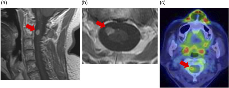
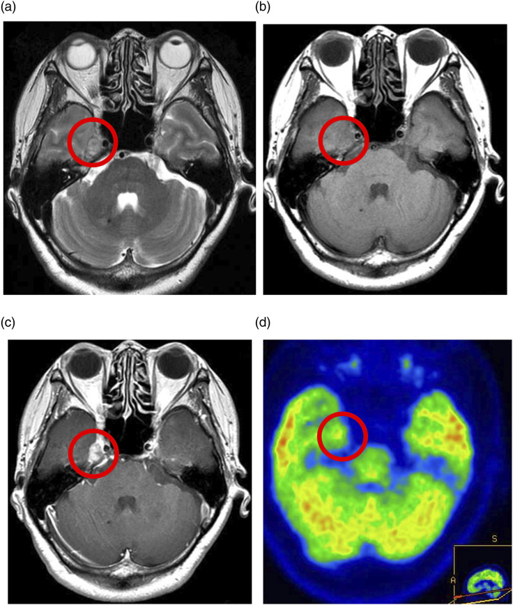
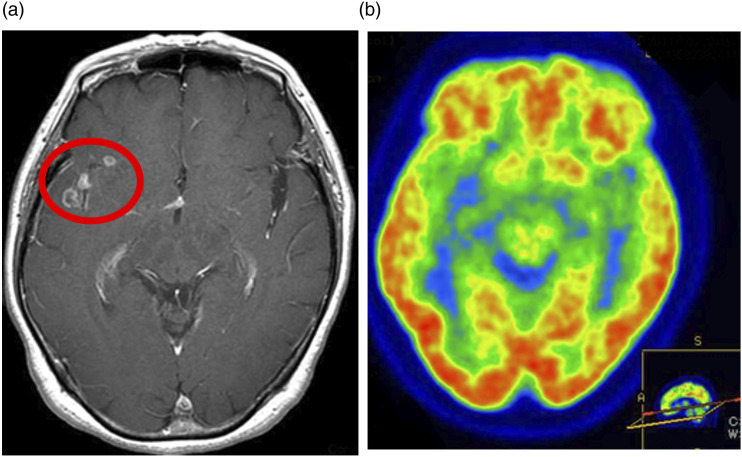
Meckel's cave or the trigeminal cistern is a subarachnoid space near the apex of the petrous portion of the temporal bone and contains cerebrospinal fluid and the Gasserian ganglion, which divides into the ophthalmic (V1), maxillary (V2), and mandibular (V3) nerves. Infectious, inflammatory, congenital, and neoplastic lesions can occur in Meckel's cave. Leptomeningeal metastasis of glioblastoma (GBM), IDH-wildtype to Meckel's cave is rare. We encountered a case of leptomeningeal metastasis of GBM to Meckel's cave in an elderly female patient who presented with pain around her right eye. Magnetic resonance imaging revealed enhancing lesions in the right temporal lobe and cervical spinal cord. The pathological diagnosis of GBM was confirmed after biopsy of the cervical spinal cord lesion, which showed hyperaccumulation of fluorodeoxyglucose (FDG) on FDG-positron emission tomography. This case indicates that metastatic lesions can also occur in Meckel's cave.


 分享
分享

 求助内容:
求助内容: 应助结果提醒方式:
 扫码关注我们
