Buyun Tian, Maoge Zhou, Fengping Feng, Xiaojun Xu, Pei Wang, Huiqin Luan, Wei Ji, Yanhong Xue, Tao Xu
{"title":"玻璃体切片的低温超分辨相关光学和电子显微镜。","authors":"Buyun Tian, Maoge Zhou, Fengping Feng, Xiaojun Xu, Pei Wang, Huiqin Luan, Wei Ji, Yanhong Xue, Tao Xu","doi":"10.52601/bpr.2022.220005","DOIUrl":null,"url":null,"abstract":"<p><p>Fluorescence microscopy and electron microscopy complement each other as the former provides labelling and localisation of specific molecules and target structures while the latter possesses excellent revolving power of fine structure in context. These two techniques can combine as correlative light and electron microscopy (CLEM) to reveal the organisation of materials within the cell. Frozen hydrated sections allow microscopic observations of cellular components <i>in situ</i> in a near-native state and are compatible with superresolution fluorescence microscopy and electron tomography if sufficient hardware and software support is available and a well-designed protocol is followed. The development of superresolution fluorescence microscopy greatly increases the precision of fluorescence annotation of electron tomograms. Here, we provide detailed instructions on how to perform cryogenic superresolution CLEM on vitreous sections. From fluorescence-labelled cells to high pressure freezing, cryo-ultramicrotomy, cryogenic single-molecule localisation microscopy, cryogenic electron tomography and image registration, electron tomograms with features of interest highlighted by superresolution fluorescence signals are expected to be obtained.</p>","PeriodicalId":59621,"journal":{"name":"生物物理学报:英文版","volume":"8 4","pages":"193-204"},"PeriodicalIF":0.0000,"publicationDate":"2022-08-31","publicationTypes":"Journal Article","fieldsOfStudy":null,"isOpenAccess":false,"openAccessPdf":"https://www.ncbi.nlm.nih.gov/pmc/articles/PMC10185487/pdf/","citationCount":"0","resultStr":"{\"title\":\"Cryogenic superresolution correlative light and electron microscopy of vitreous sections.\",\"authors\":\"Buyun Tian, Maoge Zhou, Fengping Feng, Xiaojun Xu, Pei Wang, Huiqin Luan, Wei Ji, Yanhong Xue, Tao Xu\",\"doi\":\"10.52601/bpr.2022.220005\",\"DOIUrl\":null,\"url\":null,\"abstract\":\"<p><p>Fluorescence microscopy and electron microscopy complement each other as the former provides labelling and localisation of specific molecules and target structures while the latter possesses excellent revolving power of fine structure in context. These two techniques can combine as correlative light and electron microscopy (CLEM) to reveal the organisation of materials within the cell. Frozen hydrated sections allow microscopic observations of cellular components <i>in situ</i> in a near-native state and are compatible with superresolution fluorescence microscopy and electron tomography if sufficient hardware and software support is available and a well-designed protocol is followed. The development of superresolution fluorescence microscopy greatly increases the precision of fluorescence annotation of electron tomograms. Here, we provide detailed instructions on how to perform cryogenic superresolution CLEM on vitreous sections. From fluorescence-labelled cells to high pressure freezing, cryo-ultramicrotomy, cryogenic single-molecule localisation microscopy, cryogenic electron tomography and image registration, electron tomograms with features of interest highlighted by superresolution fluorescence signals are expected to be obtained.</p>\",\"PeriodicalId\":59621,\"journal\":{\"name\":\"生物物理学报:英文版\",\"volume\":\"8 4\",\"pages\":\"193-204\"},\"PeriodicalIF\":0.0000,\"publicationDate\":\"2022-08-31\",\"publicationTypes\":\"Journal Article\",\"fieldsOfStudy\":null,\"isOpenAccess\":false,\"openAccessPdf\":\"https://www.ncbi.nlm.nih.gov/pmc/articles/PMC10185487/pdf/\",\"citationCount\":\"0\",\"resultStr\":null,\"platform\":\"Semanticscholar\",\"paperid\":null,\"PeriodicalName\":\"生物物理学报:英文版\",\"FirstCategoryId\":\"1085\",\"ListUrlMain\":\"https://doi.org/10.52601/bpr.2022.220005\",\"RegionNum\":0,\"RegionCategory\":null,\"ArticlePicture\":[],\"TitleCN\":null,\"AbstractTextCN\":null,\"PMCID\":null,\"EPubDate\":\"\",\"PubModel\":\"\",\"JCR\":\"\",\"JCRName\":\"\",\"Score\":null,\"Total\":0}","platform":"Semanticscholar","paperid":null,"PeriodicalName":"生物物理学报:英文版","FirstCategoryId":"1085","ListUrlMain":"https://doi.org/10.52601/bpr.2022.220005","RegionNum":0,"RegionCategory":null,"ArticlePicture":[],"TitleCN":null,"AbstractTextCN":null,"PMCID":null,"EPubDate":"","PubModel":"","JCR":"","JCRName":"","Score":null,"Total":0}
Cryogenic superresolution correlative light and electron microscopy of vitreous sections.
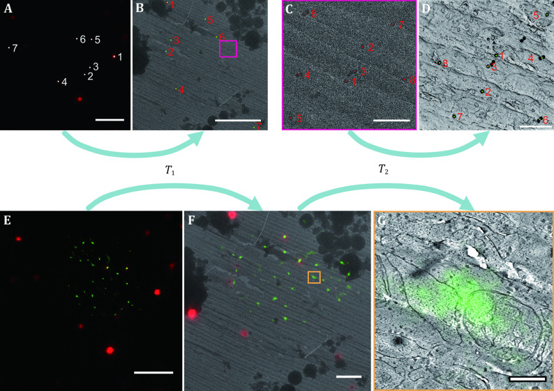
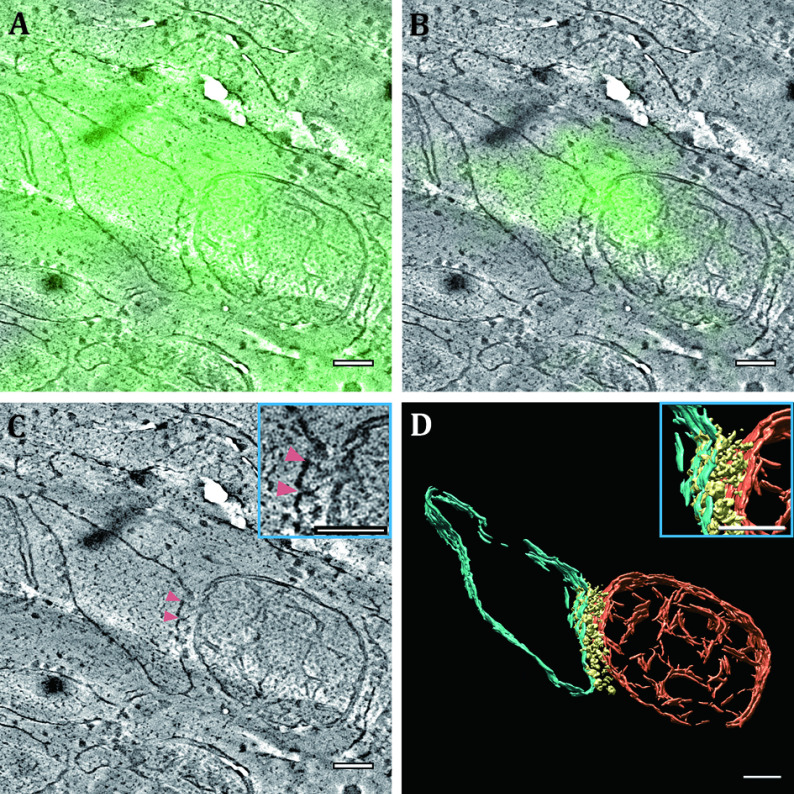
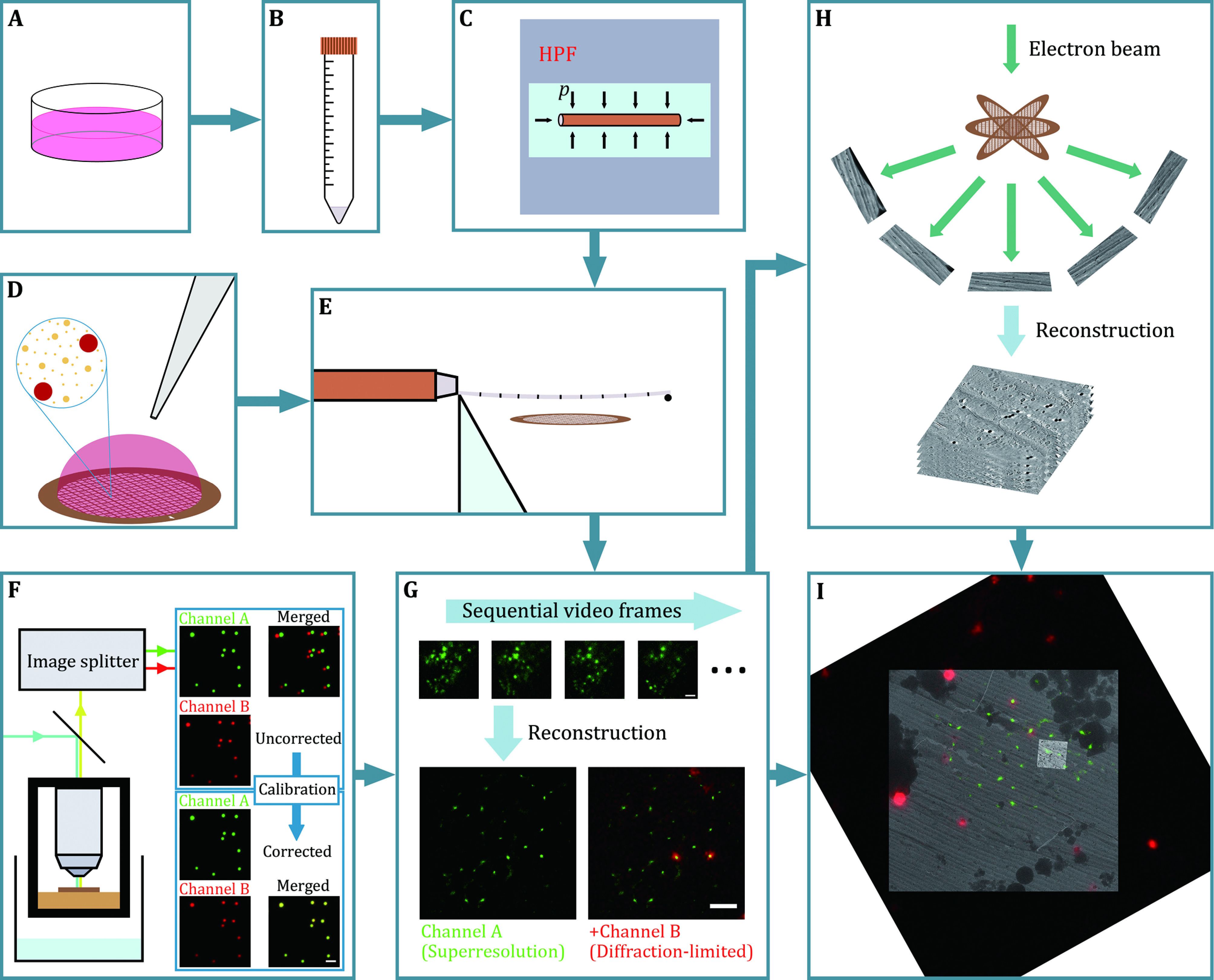
Fluorescence microscopy and electron microscopy complement each other as the former provides labelling and localisation of specific molecules and target structures while the latter possesses excellent revolving power of fine structure in context. These two techniques can combine as correlative light and electron microscopy (CLEM) to reveal the organisation of materials within the cell. Frozen hydrated sections allow microscopic observations of cellular components in situ in a near-native state and are compatible with superresolution fluorescence microscopy and electron tomography if sufficient hardware and software support is available and a well-designed protocol is followed. The development of superresolution fluorescence microscopy greatly increases the precision of fluorescence annotation of electron tomograms. Here, we provide detailed instructions on how to perform cryogenic superresolution CLEM on vitreous sections. From fluorescence-labelled cells to high pressure freezing, cryo-ultramicrotomy, cryogenic single-molecule localisation microscopy, cryogenic electron tomography and image registration, electron tomograms with features of interest highlighted by superresolution fluorescence signals are expected to be obtained.


 分享
分享
 求助内容:
求助内容: 应助结果提醒方式:
 扫码关注我们
