Sung Il Hwang, Hyungwoo Ahn, Hak Jong Lee, Sung Il Jung
{"title":"Comparative study of standard and small transrectal transducers for prostate ultrasonography.","authors":"Sung Il Hwang, Hyungwoo Ahn, Hak Jong Lee, Sung Il Jung","doi":"10.14366/usg.23084","DOIUrl":null,"url":null,"abstract":"<p><strong>Purpose: </strong>The objective of this study was to evaluate pain and image quality associated with the use of two different ultrasound transducers.</p><p><strong>Methods: </strong>Fifty healthy male participants aged 30 years or older were prospectively enrolled. All ultrasound procedures were performed using a V8 machine (Samsung Medison, Seoul, Korea) equipped with EA2-11 (conventional) and miniER7 (small-caliber) transrectal transducers, operated by a single genitourinary radiologist. To minimize bias, one group of volunteers underwent ultrasonography with the conventional transducer first, followed by the small transducer. For the remaining participants, the examinations were performed in the opposite order. Ultrasonography, including the measurement of total prostate and transitional zone volumes, was conducted in accordance with standard practice. After testing with both probes, participants were asked to rate their pain on a 10-point numerical rating scale (NRS). A radiologist then evaluated the quality of the images acquired with each probe using a 5-point numeric scale and compared the prostate volume measurements obtained by each method.</p><p><strong>Results: </strong>The mean NRS scores associated with the conventional and small transducers were 4.7±1.8 and 2.7±1.2, respectively (P<0.05). The mean ultrasound image qualities from the two transducers were statistically similar (4.78 and 4.74, P>0.05). The whole prostate gland volume as measured with the conventional transducer (mean±standard deviation, 24.2±9.1 mL) was greater than the measurement (22.1±8.7 mL) obtained with the small-caliber transducer (P<0.05). However, only two of the 50 whole gland volume measurements differed by more than two standard deviations.</p><p><strong>Conclusion: </strong>The use of a small transrectal probe significantly reduced pain without compromising image quality.</p>","PeriodicalId":54227,"journal":{"name":"Ultrasonography","volume":" ","pages":"555-560"},"PeriodicalIF":2.5000,"publicationDate":"2023-10-01","publicationTypes":"Journal Article","fieldsOfStudy":null,"isOpenAccess":false,"openAccessPdf":"https://ftp.ncbi.nlm.nih.gov/pub/pmc/oa_pdf/a3/81/usg-23084.PMC10555692.pdf","citationCount":"0","resultStr":null,"platform":"Semanticscholar","paperid":null,"PeriodicalName":"Ultrasonography","FirstCategoryId":"3","ListUrlMain":"https://doi.org/10.14366/usg.23084","RegionNum":3,"RegionCategory":"医学","ArticlePicture":[],"TitleCN":null,"AbstractTextCN":null,"PMCID":null,"EPubDate":"2023/6/30 0:00:00","PubModel":"Epub","JCR":"Q2","JCRName":"RADIOLOGY, NUCLEAR MEDICINE & MEDICAL IMAGING","Score":null,"Total":0}
引用次数: 0
Abstract
Purpose: The objective of this study was to evaluate pain and image quality associated with the use of two different ultrasound transducers.
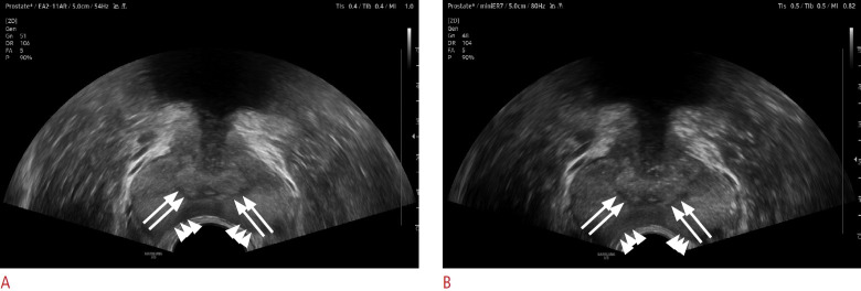
Methods: Fifty healthy male participants aged 30 years or older were prospectively enrolled. All ultrasound procedures were performed using a V8 machine (Samsung Medison, Seoul, Korea) equipped with EA2-11 (conventional) and miniER7 (small-caliber) transrectal transducers, operated by a single genitourinary radiologist. To minimize bias, one group of volunteers underwent ultrasonography with the conventional transducer first, followed by the small transducer. For the remaining participants, the examinations were performed in the opposite order. Ultrasonography, including the measurement of total prostate and transitional zone volumes, was conducted in accordance with standard practice. After testing with both probes, participants were asked to rate their pain on a 10-point numerical rating scale (NRS). A radiologist then evaluated the quality of the images acquired with each probe using a 5-point numeric scale and compared the prostate volume measurements obtained by each method.
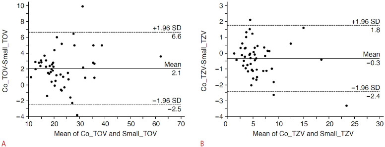
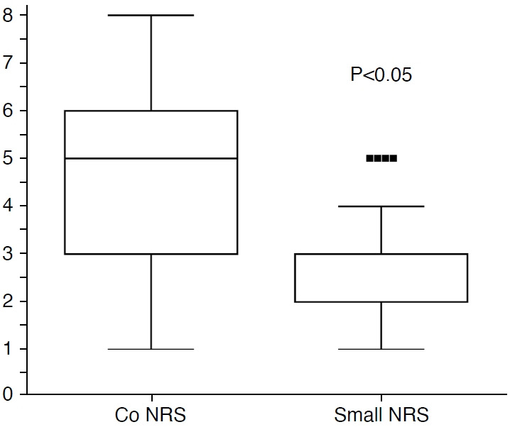
Results: The mean NRS scores associated with the conventional and small transducers were 4.7±1.8 and 2.7±1.2, respectively (P<0.05). The mean ultrasound image qualities from the two transducers were statistically similar (4.78 and 4.74, P>0.05). The whole prostate gland volume as measured with the conventional transducer (mean±standard deviation, 24.2±9.1 mL) was greater than the measurement (22.1±8.7 mL) obtained with the small-caliber transducer (P<0.05). However, only two of the 50 whole gland volume measurements differed by more than two standard deviations.
Conclusion: The use of a small transrectal probe significantly reduced pain without compromising image quality.
UltrasonographyMedicine-Radiology, Nuclear Medicine and Imaging
CiteScore
5.10
自引率
6.50%
发文量
78
审稿时长
15 weeks
期刊介绍:
Ultrasonography, the official English-language journal of the Korean Society of Ultrasound in Medicine (KSUM), is an international peer-reviewed academic journal dedicated to practice, research, technology, and education dealing with medical ultrasound. It is renamed from the Journal of Korean Society of Ultrasound in Medicine in January 2014, and published four times per year: January 1, April 1, July 1, and October 1. Original articles, technical notes, topical reviews, perspectives, pictorial essays, and timely editorial materials are published in Ultrasonography covering state-of-the-art content.
Ultrasonography aims to provide updated information on new diagnostic concepts and technical developments, including experimental animal studies using new equipment in addition to well-designed reviews of contemporary issues in patient care. Along with running KSUM Open, the annual international congress of KSUM, Ultrasonography also serves as a medium for cooperation among physicians and specialists from around the world who are focusing on various ultrasound technology and disease problems and relevant basic science.


 分享
分享
 求助内容:
求助内容: 应助结果提醒方式:
 扫码关注我们
