A M Rajani, U A Shah, Ars Mittal, S Gupta, R Garg, A A Rajani, M Punamiya, R Singhal
{"title":"AMR Sign - An Arthroscopic S-shaped Fold Signifying Adequate Medial Meniscus Repair.","authors":"A M Rajani, U A Shah, Ars Mittal, S Gupta, R Garg, A A Rajani, M Punamiya, R Singhal","doi":"10.5704/MOJ.2307.003","DOIUrl":null,"url":null,"abstract":"<p><strong>Introduction: </strong>The preferred management of medial meniscus tears has notably moved from meniscectomies towards repair. With a higher volume of meniscal repairs being done all across the world with every passing day, the lack of an objective and definitive sign suggesting the adequacy of its repair is daunting. The purpose of our study was to introduce a unique and novel arthroscopic sign formed after adequate repair of the medial meniscus, the AMR (Adequacy of Medial meniscus Repair) sign. We hypothesised that it is not only the objective end point for repair, but can also form the indicator for excellent clinical, functional, and radiological outcome even in the long term.</p><p><strong>Materials and methods: </strong>This was a multicentric, prospective study initiated by the corresponding author, and the findings validated subsequently by the other authors. Overall, it included 804 patients of isolated medial meniscus tear operated with arthroscopic all-inside technique between January 2014 and December 2017. Patients were segregated into three groups based on whether an S-shaped curve in the free, inner edge of the medial meniscus sign was formed post-repair, lost after further tightening, or not formed upon subjective completion of repair. All the patients were followed-up and evaluated based of medial joint line tenderness, McMurray's test for medial meniscus, IKDC score, WOMET score, and radiologically using an MRI at the terminal follow-up.</p><p><strong>Results: </strong>The mean terminal follow-up was 42.34±4.54 months. There was significant (p<0.01) improvement in all patients at the terminal follow-up post-surgery, irrespective of the group. The group in which AMR sign was formed and maintained showed a significantly better functional outcome on terminal follow-up as well as lower failure rates compared to the other two groups.</p><p><strong>Conclusion: </strong>AMR sign is an S-shaped fold at the inner, free edge of medial meniscus, formed after an adequate repair of isolated medial meniscus tear, as viewed on arthroscopy. It is an objective sign denoting regained integrity of the collagen architecture of the medial meniscus following repair. It is also a reliable indicator of excellent long term functional, clinical, and radiological outcome and also lower failure rates in patients after arthroscopic medial meniscus repair.</p>","PeriodicalId":45241,"journal":{"name":"Malaysian Orthopaedic Journal","volume":"17 2","pages":"13-20"},"PeriodicalIF":0.6000,"publicationDate":"2023-07-01","publicationTypes":"Journal Article","fieldsOfStudy":null,"isOpenAccess":false,"openAccessPdf":"https://www.ncbi.nlm.nih.gov/pmc/articles/PMC10425007/pdf/","citationCount":"0","resultStr":null,"platform":"Semanticscholar","paperid":null,"PeriodicalName":"Malaysian Orthopaedic Journal","FirstCategoryId":"1085","ListUrlMain":"https://doi.org/10.5704/MOJ.2307.003","RegionNum":0,"RegionCategory":null,"ArticlePicture":[],"TitleCN":null,"AbstractTextCN":null,"PMCID":null,"EPubDate":"","PubModel":"","JCR":"Q4","JCRName":"ORTHOPEDICS","Score":null,"Total":0}
引用次数: 0
Abstract
Introduction: The preferred management of medial meniscus tears has notably moved from meniscectomies towards repair. With a higher volume of meniscal repairs being done all across the world with every passing day, the lack of an objective and definitive sign suggesting the adequacy of its repair is daunting. The purpose of our study was to introduce a unique and novel arthroscopic sign formed after adequate repair of the medial meniscus, the AMR (Adequacy of Medial meniscus Repair) sign. We hypothesised that it is not only the objective end point for repair, but can also form the indicator for excellent clinical, functional, and radiological outcome even in the long term.
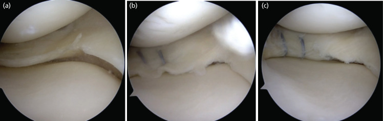
Materials and methods: This was a multicentric, prospective study initiated by the corresponding author, and the findings validated subsequently by the other authors. Overall, it included 804 patients of isolated medial meniscus tear operated with arthroscopic all-inside technique between January 2014 and December 2017. Patients were segregated into three groups based on whether an S-shaped curve in the free, inner edge of the medial meniscus sign was formed post-repair, lost after further tightening, or not formed upon subjective completion of repair. All the patients were followed-up and evaluated based of medial joint line tenderness, McMurray's test for medial meniscus, IKDC score, WOMET score, and radiologically using an MRI at the terminal follow-up.
Results: The mean terminal follow-up was 42.34±4.54 months. There was significant (p<0.01) improvement in all patients at the terminal follow-up post-surgery, irrespective of the group. The group in which AMR sign was formed and maintained showed a significantly better functional outcome on terminal follow-up as well as lower failure rates compared to the other two groups.
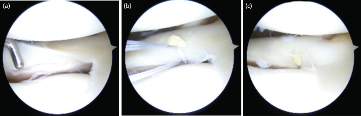
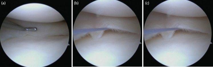
Conclusion: AMR sign is an S-shaped fold at the inner, free edge of medial meniscus, formed after an adequate repair of isolated medial meniscus tear, as viewed on arthroscopy. It is an objective sign denoting regained integrity of the collagen architecture of the medial meniscus following repair. It is also a reliable indicator of excellent long term functional, clinical, and radiological outcome and also lower failure rates in patients after arthroscopic medial meniscus repair.
期刊介绍:
The Malaysian Orthopaedic Journal is a peer-reviewed journal that publishes original papers and case reports three times a year in both printed and electronic version. The purpose of MOJ is to disseminate new knowledge and provide updates in Orthopaedics, trauma and musculoskeletal research. It is an Open Access journal that does not require processing fee or article processing charge from the authors. The Malaysian Orthopaedic Journal is the official journal of Malaysian Orthopaedic Association (MOA) and ASEAN Orthopaedic Association (AOA).


 分享
分享
 求助内容:
求助内容: 应助结果提醒方式:
 扫码关注我们
