Daniele Bertoglio, Alison R Weiss, William Liguore, Lauren Drew Martin, Theodore Hobbs, John Templon, Sathya Srinivasan, Celia Dominguez, Ignacio Munoz-Sanjuan, Vinod Khetarpal, Jeroen Verhaeghe, Steven Staelens, Jeanne Link, Longbin Liu, Jonathan A Bard, Jodi L McBride
{"title":"In Vivo Cerebral Imaging of Mutant Huntingtin Aggregates Using <sup>11</sup>C-CHDI-180R PET in a Nonhuman Primate Model of Huntington Disease.","authors":"Daniele Bertoglio, Alison R Weiss, William Liguore, Lauren Drew Martin, Theodore Hobbs, John Templon, Sathya Srinivasan, Celia Dominguez, Ignacio Munoz-Sanjuan, Vinod Khetarpal, Jeroen Verhaeghe, Steven Staelens, Jeanne Link, Longbin Liu, Jonathan A Bard, Jodi L McBride","doi":"10.2967/jnumed.123.265569","DOIUrl":null,"url":null,"abstract":"<p><p>Huntington disease (HD) is a neurodegenerative disorder caused by an expanded polyglutamine (CAG) trinucleotide expansion in the huntingtin (<i>HTT</i>) gene that encodes the mutant huntingtin protein (mHTT). Visualization and quantification of cerebral mHTT will provide a proxy for target engagement and a means to evaluate therapeutic interventions aimed at lowering mHTT in the brain. Here, we validated the novel radioligand <sup>11</sup>C-labeled 6-(5-((5-methoxypyridin-2-yl)methoxy)benzo[d]oxazol-2-yl)-2-methylpyridazin-3(2H)-one (<sup>11</sup>C-CHDI-180R) using PET imaging to quantify cerebral mHTT aggregates in a macaque model of HD. <b>Methods:</b> Rhesus macaques received MRI-guided intrastriatal delivery of a mixture of AAV2 and AAV2.retro viral vectors expressing an HTT fragment bearing 85 CAG repeats (85Q, <i>n</i> = 5), a control HTT fragment bearing 10 CAG repeats (10Q, <i>n</i> = 4), or vector diluent only (phosphate-buffered saline, <i>n</i> = 5). Thirty months after surgery, 90-min dynamic PET/CT imaging was used to investigate <sup>11</sup>C-CHDI-180R brain kinetics, along with serial blood sampling to measure input function and stability of the radioligand. The total volume of distribution was calculated using a 2-tissue-compartment model as well as Logan graphical analysis for regional quantification. Immunostaining for mHTT was performed to corroborate the in vivo findings. <b>Results:</b> <sup>11</sup>C-CHDI-180R displayed good metabolic stability (51.4% ± 4.0% parent in plasma at 60 min after injection). Regional time-activity curves displayed rapid uptake and reversible binding, which were described by a 2-tissue-compartment model. Logan graphical analysis was associated with the 2-tissue-compartment model (<i>r</i> <sup>2</sup> = 0.96, <i>P</i> < 0.0001) and used to generate parametric volume of distribution maps. Compared with controls, animals administered the 85Q fragment exhibited significantly increased <sup>11</sup>C-CHDI-180R binding in several cortical and subcortical brain regions (group effect, <i>P</i> < 0.0001). No difference in <sup>11</sup>C-CHDI-180R binding was observed between buffer and 10Q animals. The presence of mHTT aggregates in the 85Q animals was confirmed histologically. <b>Conclusion:</b> We validated <sup>11</sup>C-CHDI-180R as a radioligand to visualize and quantify mHTT aggregated species in a HD macaque model. These findings corroborate our previous work in rodent HD models and show that <sup>11</sup>C-CHDI-180R is a promising tool to assess the mHTT aggregate load and the efficacy of therapeutic strategies.</p>","PeriodicalId":16758,"journal":{"name":"Journal of Nuclear Medicine","volume":" ","pages":"1581-1587"},"PeriodicalIF":9.1000,"publicationDate":"2023-10-01","publicationTypes":"Journal Article","fieldsOfStudy":null,"isOpenAccess":false,"openAccessPdf":"https://www.ncbi.nlm.nih.gov/pmc/articles/PMC10586486/pdf/","citationCount":"0","resultStr":null,"platform":"Semanticscholar","paperid":null,"PeriodicalName":"Journal of Nuclear Medicine","FirstCategoryId":"3","ListUrlMain":"https://doi.org/10.2967/jnumed.123.265569","RegionNum":1,"RegionCategory":"医学","ArticlePicture":[],"TitleCN":null,"AbstractTextCN":null,"PMCID":null,"EPubDate":"2023/8/17 0:00:00","PubModel":"Epub","JCR":"Q1","JCRName":"RADIOLOGY, NUCLEAR MEDICINE & MEDICAL IMAGING","Score":null,"Total":0}
引用次数: 0
Abstract
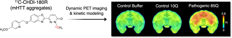
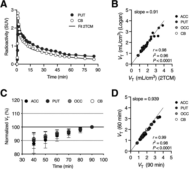
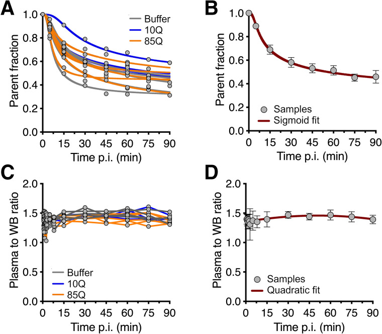
Huntington disease (HD) is a neurodegenerative disorder caused by an expanded polyglutamine (CAG) trinucleotide expansion in the huntingtin (HTT) gene that encodes the mutant huntingtin protein (mHTT). Visualization and quantification of cerebral mHTT will provide a proxy for target engagement and a means to evaluate therapeutic interventions aimed at lowering mHTT in the brain. Here, we validated the novel radioligand 11C-labeled 6-(5-((5-methoxypyridin-2-yl)methoxy)benzo[d]oxazol-2-yl)-2-methylpyridazin-3(2H)-one (11C-CHDI-180R) using PET imaging to quantify cerebral mHTT aggregates in a macaque model of HD. Methods: Rhesus macaques received MRI-guided intrastriatal delivery of a mixture of AAV2 and AAV2.retro viral vectors expressing an HTT fragment bearing 85 CAG repeats (85Q, n = 5), a control HTT fragment bearing 10 CAG repeats (10Q, n = 4), or vector diluent only (phosphate-buffered saline, n = 5). Thirty months after surgery, 90-min dynamic PET/CT imaging was used to investigate 11C-CHDI-180R brain kinetics, along with serial blood sampling to measure input function and stability of the radioligand. The total volume of distribution was calculated using a 2-tissue-compartment model as well as Logan graphical analysis for regional quantification. Immunostaining for mHTT was performed to corroborate the in vivo findings. Results:11C-CHDI-180R displayed good metabolic stability (51.4% ± 4.0% parent in plasma at 60 min after injection). Regional time-activity curves displayed rapid uptake and reversible binding, which were described by a 2-tissue-compartment model. Logan graphical analysis was associated with the 2-tissue-compartment model (r2 = 0.96, P < 0.0001) and used to generate parametric volume of distribution maps. Compared with controls, animals administered the 85Q fragment exhibited significantly increased 11C-CHDI-180R binding in several cortical and subcortical brain regions (group effect, P < 0.0001). No difference in 11C-CHDI-180R binding was observed between buffer and 10Q animals. The presence of mHTT aggregates in the 85Q animals was confirmed histologically. Conclusion: We validated 11C-CHDI-180R as a radioligand to visualize and quantify mHTT aggregated species in a HD macaque model. These findings corroborate our previous work in rodent HD models and show that 11C-CHDI-180R is a promising tool to assess the mHTT aggregate load and the efficacy of therapeutic strategies.
期刊介绍:
The Journal of Nuclear Medicine (JNM), self-published by the Society of Nuclear Medicine and Molecular Imaging (SNMMI), provides readers worldwide with clinical and basic science investigations, continuing education articles, reviews, employment opportunities, and updates on practice and research. In the 2022 Journal Citation Reports (released in June 2023), JNM ranked sixth in impact among 203 medical journals worldwide in the radiology, nuclear medicine, and medical imaging category.


 分享
分享
 求助内容:
求助内容: 应助结果提醒方式:
 扫码关注我们
