Retta El Sayed, Alireza Sharifi, Charlie C Park, Diogo C Haussen, Jason W Allen, John N Oshinski
{"title":"Optimization of 4D Flow MRI Spatial and Temporal Resolution for Examining Complex Hemodynamics in the Carotid Artery Bifurcation.","authors":"Retta El Sayed, Alireza Sharifi, Charlie C Park, Diogo C Haussen, Jason W Allen, John N Oshinski","doi":"10.1007/s13239-023-00667-1","DOIUrl":null,"url":null,"abstract":"<p><strong>Background: </strong>Three-dimensional, ECG-gated, time-resolved, three-directional, velocity-encoded phase-contrast MRI (4D flow MRI) has been applied extensively to measure blood velocity in great vessels but has been much less used in diseased carotid arteries. Carotid artery webs (CaW) are non-inflammatory intraluminal shelf-like projections into the internal carotid artery (ICA) bulb that are associated with complex flow and cryptogenic stroke.</p><p><strong>Purpose: </strong>Optimize 4D flow MRI for measuring the velocity field of complex flow in the carotid artery bifurcation model that contains a CaW.</p><p><strong>Methods: </strong>A 3D printed phantom model created from computed tomography angiography (CTA) of a subject with CaW was placed in a pulsatile flow loop within the MRI scanner. 4D Flow MRI images of the phantom were acquired with five different spatial resolutions (0.50-2.00 mm<sup>3</sup>) and four different temporal resolutions (23-96 ms) and compared to a computational fluid dynamics (CFD) solution of the flow field as a reference. We examined four planes perpendicular to the vessel centerline, one in the common carotid artery (CCA) and three in the internal carotid artery (ICA) where complex flow was expected. At these four planes pixel-by-pixel velocity values, flow, and time average wall shear stress (TAWSS) were compared between 4D flow MRI and CFD.</p><p><strong>Hypothesis: </strong>An optimized 4D flow MRI protocol will provide a good correlation with CFD velocity and TAWSS values in areas of complex flow within a clinically feasible scan time (~ 10 min).</p><p><strong>Results: </strong>Spatial resolution affected the velocity values, time average flow, and TAWSS measurements. Qualitatively, a spatial resolution of 0.50 mm<sup>3</sup> resulted in higher noise, while a lower spatial resolution of 1.50-2.00 mm<sup>3</sup> did not adequately resolve the velocity profile. Isotropic spatial resolutions of 0.50-1.00 mm<sup>3</sup> showed no significant difference in total flow compared to CFD. Pixel-by-pixel velocity correlation coefficients between 4D flow MRI and CFD were > 0.75 for 0.50-1.00 mm<sup>3</sup> but were < 0.5 for 1.50 and 2.00 mm<sup>3</sup>. Regional TAWSS values determined from 4D flow MRI were generally lower than CFD and decreased at lower spatial resolutions (larger pixel sizes). TAWSS differences between 4D flow and CFD were not statistically significant at spatial resolutions of 0.50-1.00 mm<sup>3</sup> but were different at 1.50 and 2.00 mm<sup>3</sup>. Differences in temporal resolution only affected the flow values when temporal resolution was > 48.4 ms; temporal resolution did not affect TAWSS values.</p><p><strong>Conclusion: </strong>A spatial resolution of 0.74-1.00 mm<sup>3</sup> and a temporal resolution of 23-48 ms (1-2 k-space segments) provides a 4D flow MRI protocol capable of imaging velocity and TAWSS in regions of complex flow within the carotid bifurcation at a clinically acceptable scan time.</p>","PeriodicalId":54322,"journal":{"name":"Cardiovascular Engineering and Technology","volume":"14 3","pages":"476-488"},"PeriodicalIF":1.8000,"publicationDate":"2023-06-01","publicationTypes":"Journal Article","fieldsOfStudy":null,"isOpenAccess":false,"openAccessPdf":"https://www.ncbi.nlm.nih.gov/pmc/articles/PMC10524741/pdf/nihms-1901196.pdf","citationCount":"1","resultStr":null,"platform":"Semanticscholar","paperid":null,"PeriodicalName":"Cardiovascular Engineering and Technology","FirstCategoryId":"5","ListUrlMain":"https://doi.org/10.1007/s13239-023-00667-1","RegionNum":4,"RegionCategory":"医学","ArticlePicture":[],"TitleCN":null,"AbstractTextCN":null,"PMCID":null,"EPubDate":"2023/5/8 0:00:00","PubModel":"Epub","JCR":"Q3","JCRName":"CARDIAC & CARDIOVASCULAR SYSTEMS","Score":null,"Total":0}
引用次数: 1
Abstract
Background: Three-dimensional, ECG-gated, time-resolved, three-directional, velocity-encoded phase-contrast MRI (4D flow MRI) has been applied extensively to measure blood velocity in great vessels but has been much less used in diseased carotid arteries. Carotid artery webs (CaW) are non-inflammatory intraluminal shelf-like projections into the internal carotid artery (ICA) bulb that are associated with complex flow and cryptogenic stroke.
Purpose: Optimize 4D flow MRI for measuring the velocity field of complex flow in the carotid artery bifurcation model that contains a CaW.
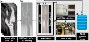
Methods: A 3D printed phantom model created from computed tomography angiography (CTA) of a subject with CaW was placed in a pulsatile flow loop within the MRI scanner. 4D Flow MRI images of the phantom were acquired with five different spatial resolutions (0.50-2.00 mm3) and four different temporal resolutions (23-96 ms) and compared to a computational fluid dynamics (CFD) solution of the flow field as a reference. We examined four planes perpendicular to the vessel centerline, one in the common carotid artery (CCA) and three in the internal carotid artery (ICA) where complex flow was expected. At these four planes pixel-by-pixel velocity values, flow, and time average wall shear stress (TAWSS) were compared between 4D flow MRI and CFD.
Hypothesis: An optimized 4D flow MRI protocol will provide a good correlation with CFD velocity and TAWSS values in areas of complex flow within a clinically feasible scan time (~ 10 min).
Results: Spatial resolution affected the velocity values, time average flow, and TAWSS measurements. Qualitatively, a spatial resolution of 0.50 mm3 resulted in higher noise, while a lower spatial resolution of 1.50-2.00 mm3 did not adequately resolve the velocity profile. Isotropic spatial resolutions of 0.50-1.00 mm3 showed no significant difference in total flow compared to CFD. Pixel-by-pixel velocity correlation coefficients between 4D flow MRI and CFD were > 0.75 for 0.50-1.00 mm3 but were < 0.5 for 1.50 and 2.00 mm3. Regional TAWSS values determined from 4D flow MRI were generally lower than CFD and decreased at lower spatial resolutions (larger pixel sizes). TAWSS differences between 4D flow and CFD were not statistically significant at spatial resolutions of 0.50-1.00 mm3 but were different at 1.50 and 2.00 mm3. Differences in temporal resolution only affected the flow values when temporal resolution was > 48.4 ms; temporal resolution did not affect TAWSS values.
Conclusion: A spatial resolution of 0.74-1.00 mm3 and a temporal resolution of 23-48 ms (1-2 k-space segments) provides a 4D flow MRI protocol capable of imaging velocity and TAWSS in regions of complex flow within the carotid bifurcation at a clinically acceptable scan time.
期刊介绍:
Cardiovascular Engineering and Technology is a journal publishing the spectrum of basic to translational research in all aspects of cardiovascular physiology and medical treatment. It is the forum for academic and industrial investigators to disseminate research that utilizes engineering principles and methods to advance fundamental knowledge and technological solutions related to the cardiovascular system. Manuscripts spanning from subcellular to systems level topics are invited, including but not limited to implantable medical devices, hemodynamics and tissue biomechanics, functional imaging, surgical devices, electrophysiology, tissue engineering and regenerative medicine, diagnostic instruments, transport and delivery of biologics, and sensors. In addition to manuscripts describing the original publication of research, manuscripts reviewing developments in these topics or their state-of-art are also invited.


 分享
分享
 求助内容:
求助内容: 应助结果提醒方式:
 扫码关注我们
