{"title":"[<sup>68</sup>Ga] Ga-Pentixafor diffuse bilateral Adrenal & Breast uptake in a patient with High-grade Glioma: A note of caution on normal variants.","authors":"Hessamoddin Roustaei, Emran Askari, Somayeh Barashki, Kazem Anvari, Ramin Sadeghi, Kamran Aryana","doi":"10.22038/AOJNMB.2022.66223.1458","DOIUrl":null,"url":null,"abstract":"<p><p>[<sup>68</sup>Ga] Ga-labeled C-X-C motif receptor4 as a novel radio-ligand using PET/CT has been investigated for tracing various kinds of solid and hematopoietic malignancies in recent years. High-grade Glioma (WHO classification 2016 grade III and IV) shows elevated levels of CXCR4 ligand expression in the affected tumoral cells. Healthy and non-affected organ cells express low-level CXCR4 ligands density. We performed [<sup>68</sup>Ga] Ga-Pentixafor (Pars-Cixafor™) PET/CT in a patient with high-grade Glioma (anaplastic oligodendroglioma WHO grade III) with no other documented medical condition and history. In addition to the Pentixafor-avid tumor remnant in the PET/CT images, we observed mild symmetrical bilateral uptake in the fibro glandular tissue of the breasts and moderate CXCR4(Pentixafor) avidity in both adrenal glands without any discernable pathology and abnormal density changes in the CT component of the study. Attention should be paid to the interpreting [<sup>68</sup>Ga] Ga-Pentixafor PET/CT examination and its normal uptakes and variants.</p>","PeriodicalId":72309,"journal":{"name":"","volume":"11 2","pages":"168-170"},"PeriodicalIF":0.0,"publicationDate":"2023-01-01","publicationTypes":"Journal Article","fieldsOfStudy":null,"isOpenAccess":false,"openAccessPdf":"https://www.ncbi.nlm.nih.gov/pmc/articles/PMC10261692/pdf/","citationCount":"0","resultStr":null,"platform":"Semanticscholar","paperid":null,"PeriodicalName":"","FirstCategoryId":"1085","ListUrlMain":"https://doi.org/10.22038/AOJNMB.2022.66223.1458","RegionNum":0,"RegionCategory":null,"ArticlePicture":[],"TitleCN":null,"AbstractTextCN":null,"PMCID":null,"EPubDate":"","PubModel":"","JCR":"","JCRName":"","Score":null,"Total":0}
引用次数: 0
Abstract
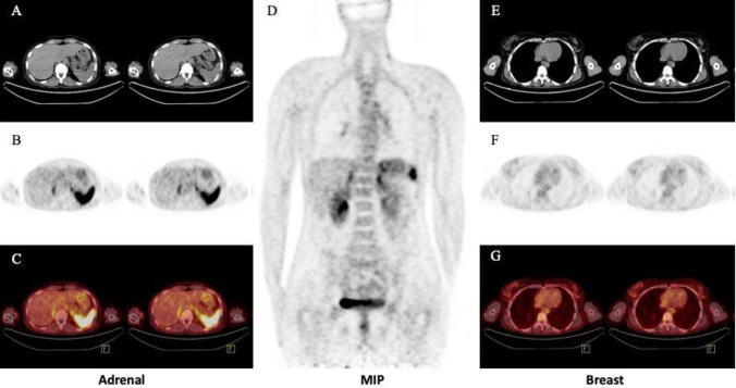
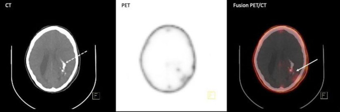
[68Ga] Ga-labeled C-X-C motif receptor4 as a novel radio-ligand using PET/CT has been investigated for tracing various kinds of solid and hematopoietic malignancies in recent years. High-grade Glioma (WHO classification 2016 grade III and IV) shows elevated levels of CXCR4 ligand expression in the affected tumoral cells. Healthy and non-affected organ cells express low-level CXCR4 ligands density. We performed [68Ga] Ga-Pentixafor (Pars-Cixafor™) PET/CT in a patient with high-grade Glioma (anaplastic oligodendroglioma WHO grade III) with no other documented medical condition and history. In addition to the Pentixafor-avid tumor remnant in the PET/CT images, we observed mild symmetrical bilateral uptake in the fibro glandular tissue of the breasts and moderate CXCR4(Pentixafor) avidity in both adrenal glands without any discernable pathology and abnormal density changes in the CT component of the study. Attention should be paid to the interpreting [68Ga] Ga-Pentixafor PET/CT examination and its normal uptakes and variants.


 分享
分享
 求助内容:
求助内容: 应助结果提醒方式:
 扫码关注我们
