Gonzalo Israel Gutiérrez-Díaz, Arturo David Buenrostro-Jiménez, Roberto Rojas-Castillo, Víctor Amador-Avendaño, Alma Yaneth Jaime-Zúñiga, Anahí de Jesús Zambada-Gamboa, Manuel Alejandro Velázquez-García, Diego Armando Gudiño-Amezcua
{"title":"[Acute coronary syndrome provoked by a coronary intramural hematoma].","authors":"Gonzalo Israel Gutiérrez-Díaz, Arturo David Buenrostro-Jiménez, Roberto Rojas-Castillo, Víctor Amador-Avendaño, Alma Yaneth Jaime-Zúñiga, Anahí de Jesús Zambada-Gamboa, Manuel Alejandro Velázquez-García, Diego Armando Gudiño-Amezcua","doi":"","DOIUrl":null,"url":null,"abstract":"<p><strong>Background: </strong>Intramural coronary hematoma (ICH) is an unusual cause of acute coronary syndrome, and it represents a diagnostic challenge, especially in young patients in which it is not considered among the differential causes of acute myocardial ischemia.</p><p><strong>Clinical case: </strong>40-year-old female, with type 2 diabetes and no other cardiovascular risk factors, who assisted to the Emergency Room with chest pain. In her first evaluation, electrocardiographic abnormalities, and troponin I elevation were found. A cardiac catheterization was performed, in which a proximal obstruction of the left anterior descending artery was observed, and then an optical coherence tomography (OCT) confirmed the presence of an ICH without a dissection flap. A stent was implanted in the obstruction area, with adequate angiographic outcome. The patient had a satisfactory evolution and was discharged to home without evidence of systolic dysfunction and is free of cardiovascular symptoms at 6-month follow-up.</p><p><strong>Conclusions: </strong>ICH must be considered within the differential diagnosis of acute myocardial ischemia in young patients, especially females. Intravascular image diagnosis is essential for the adequate diagnosis and treatment. Treatment must be individualized considering the extent of ischemia.</p>","PeriodicalId":21419,"journal":{"name":"Revista médica del Instituto Mexicano del Seguro Social","volume":"61 3","pages":"380-385"},"PeriodicalIF":0.0000,"publicationDate":"2023-05-02","publicationTypes":"Journal Article","fieldsOfStudy":null,"isOpenAccess":false,"openAccessPdf":"https://ftp.ncbi.nlm.nih.gov/pub/pmc/oa_pdf/9d/43/04435117-61-3-380.PMC10437236.pdf","citationCount":"0","resultStr":null,"platform":"Semanticscholar","paperid":null,"PeriodicalName":"Revista médica del Instituto Mexicano del Seguro Social","FirstCategoryId":"1085","ListUrlMain":"","RegionNum":0,"RegionCategory":null,"ArticlePicture":[],"TitleCN":null,"AbstractTextCN":null,"PMCID":null,"EPubDate":"","PubModel":"","JCR":"","JCRName":"","Score":null,"Total":0}
引用次数: 0
Abstract
Background: Intramural coronary hematoma (ICH) is an unusual cause of acute coronary syndrome, and it represents a diagnostic challenge, especially in young patients in which it is not considered among the differential causes of acute myocardial ischemia.
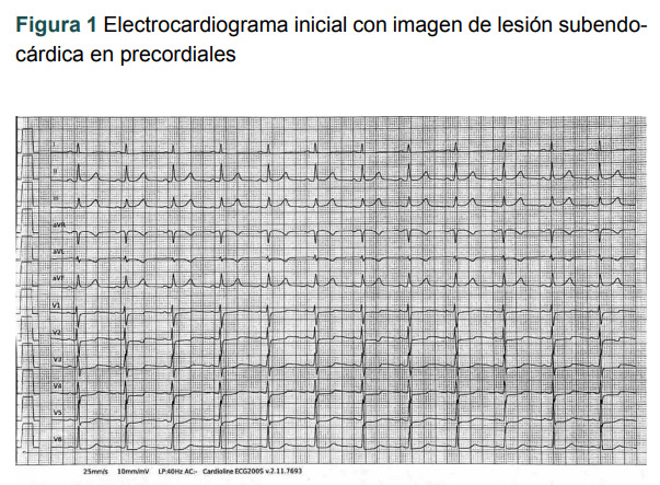
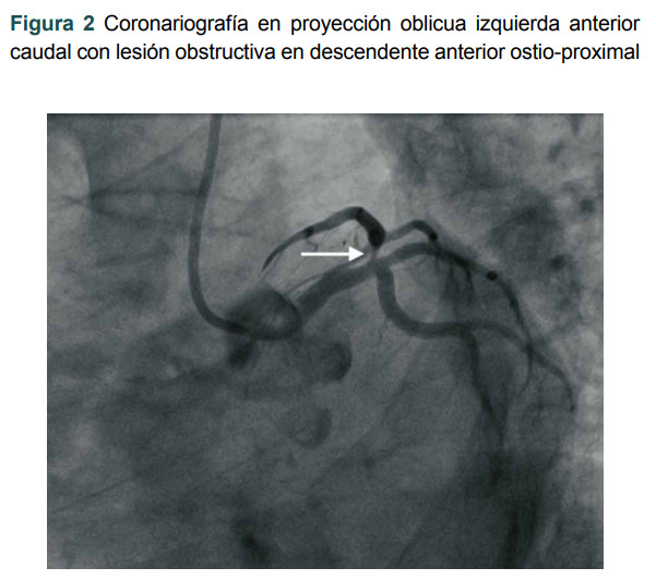
Clinical case: 40-year-old female, with type 2 diabetes and no other cardiovascular risk factors, who assisted to the Emergency Room with chest pain. In her first evaluation, electrocardiographic abnormalities, and troponin I elevation were found. A cardiac catheterization was performed, in which a proximal obstruction of the left anterior descending artery was observed, and then an optical coherence tomography (OCT) confirmed the presence of an ICH without a dissection flap. A stent was implanted in the obstruction area, with adequate angiographic outcome. The patient had a satisfactory evolution and was discharged to home without evidence of systolic dysfunction and is free of cardiovascular symptoms at 6-month follow-up.
Conclusions: ICH must be considered within the differential diagnosis of acute myocardial ischemia in young patients, especially females. Intravascular image diagnosis is essential for the adequate diagnosis and treatment. Treatment must be individualized considering the extent of ischemia.
 分享
分享


 求助内容:
求助内容: 应助结果提醒方式:
 扫码关注我们
