Fibroblast activation protein-α expression in fibroblasts is common in the tumor microenvironment of colorectal cancer and may serve as a therapeutic target.
K Greimelmaier, N Klopp, E Mairinger, M Wessolly, S Borchert, J Steinborn, K W Schmid, J Wohlschlaeger, F D Mairinger
{"title":"Fibroblast activation protein-α expression in fibroblasts is common in the tumor microenvironment of colorectal cancer and may serve as a therapeutic target.","authors":"K Greimelmaier, N Klopp, E Mairinger, M Wessolly, S Borchert, J Steinborn, K W Schmid, J Wohlschlaeger, F D Mairinger","doi":"10.3389/pore.2023.1611163","DOIUrl":null,"url":null,"abstract":"<p><p><b>Background:</b> Colorectal cancer (CRC) is still one of the leading causes of cancer death worldwide, emphasizing the need for further diagnostic and therapeutic approaches. Cancer invasion and metastasis are affected by the tumor microenvironment (TME), with cancer-associated fibroblasts (CAF) being the predominant cellular component. An important marker for CAF is fibroblast activation protein-α (FAP) which has been evaluated as therapeutic target for, e.g., radioligand therapy. The aim of this study was to examine CRC regarding the FAP expression as a candidate for targeted therapy. <b>Methods:</b> 67 CRC, 24 adenomas, 18 tissue samples of inflammation sites and 28 non-neoplastic, non-inflammatory tissue samples of colonic mucosa were evaluated for immunohistochemical FAP expression of CAF in tissue microarrays. The results were correlated with clinicopathological data, tumor biology and concurrent expression of additional immunohistochemical parameters. <b>Results:</b> 53/67 (79%) CRC and 6/18 (33%) inflammatory tissue specimens showed expression of FAP. However, FAP was only present in 1/24 (4%) adenomas and absent in normal mucosa (0/28). Thus, FAP expression in CRC was significantly higher than in the other investigated groups. Within the CRC cohort, expression of FAP did not correlate with tumor stage, grading or the MSI status. However, it was observed that tumors exhibiting high immunohistochemical expression of Ki-67, CD3, p53, and β-Catenin showed a significantly higher incidence of FAP expression. <b>Conclusion:</b> In the crosstalk between tumor cells and TME, CAF play a key role in carcinogenesis and metastatic spread. Expression of FAP was detectable in the majority of CRC but nearly absent in precursor lesions and non-neoplastic, non-inflammatory tissue. This finding indicates that FAP has the potential to emerge as a target for new diagnostic and therapeutic concepts in CRC. Additionally, the association between FAP expression and other immunohistochemical parameters displays the interaction between different components of the TME and demands further investigation.</p>","PeriodicalId":19981,"journal":{"name":"Pathology & Oncology Research","volume":"29 ","pages":"1611163"},"PeriodicalIF":2.3000,"publicationDate":"2023-01-01","publicationTypes":"Journal Article","fieldsOfStudy":null,"isOpenAccess":false,"openAccessPdf":"https://www.ncbi.nlm.nih.gov/pmc/articles/PMC10442481/pdf/","citationCount":"1","resultStr":null,"platform":"Semanticscholar","paperid":null,"PeriodicalName":"Pathology & Oncology Research","FirstCategoryId":"3","ListUrlMain":"https://doi.org/10.3389/pore.2023.1611163","RegionNum":4,"RegionCategory":"医学","ArticlePicture":[],"TitleCN":null,"AbstractTextCN":null,"PMCID":null,"EPubDate":"","PubModel":"","JCR":"Q3","JCRName":"ONCOLOGY","Score":null,"Total":0}
引用次数: 1
Abstract
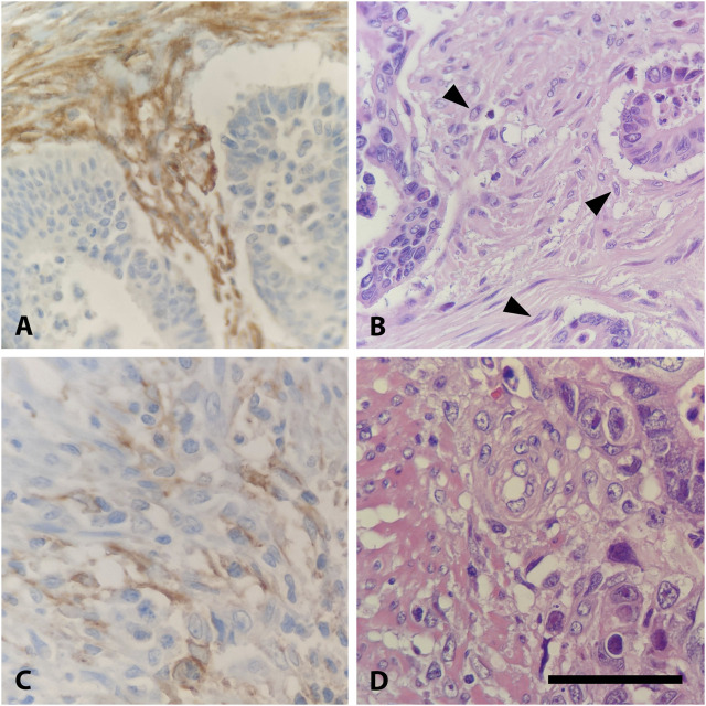
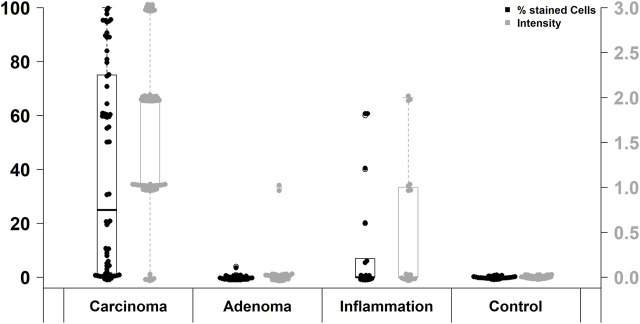
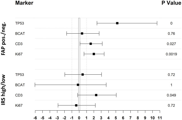
Background: Colorectal cancer (CRC) is still one of the leading causes of cancer death worldwide, emphasizing the need for further diagnostic and therapeutic approaches. Cancer invasion and metastasis are affected by the tumor microenvironment (TME), with cancer-associated fibroblasts (CAF) being the predominant cellular component. An important marker for CAF is fibroblast activation protein-α (FAP) which has been evaluated as therapeutic target for, e.g., radioligand therapy. The aim of this study was to examine CRC regarding the FAP expression as a candidate for targeted therapy. Methods: 67 CRC, 24 adenomas, 18 tissue samples of inflammation sites and 28 non-neoplastic, non-inflammatory tissue samples of colonic mucosa were evaluated for immunohistochemical FAP expression of CAF in tissue microarrays. The results were correlated with clinicopathological data, tumor biology and concurrent expression of additional immunohistochemical parameters. Results: 53/67 (79%) CRC and 6/18 (33%) inflammatory tissue specimens showed expression of FAP. However, FAP was only present in 1/24 (4%) adenomas and absent in normal mucosa (0/28). Thus, FAP expression in CRC was significantly higher than in the other investigated groups. Within the CRC cohort, expression of FAP did not correlate with tumor stage, grading or the MSI status. However, it was observed that tumors exhibiting high immunohistochemical expression of Ki-67, CD3, p53, and β-Catenin showed a significantly higher incidence of FAP expression. Conclusion: In the crosstalk between tumor cells and TME, CAF play a key role in carcinogenesis and metastatic spread. Expression of FAP was detectable in the majority of CRC but nearly absent in precursor lesions and non-neoplastic, non-inflammatory tissue. This finding indicates that FAP has the potential to emerge as a target for new diagnostic and therapeutic concepts in CRC. Additionally, the association between FAP expression and other immunohistochemical parameters displays the interaction between different components of the TME and demands further investigation.
期刊介绍:
Pathology & Oncology Research (POR) is an interdisciplinary Journal at the interface of pathology and oncology including the preclinical and translational research, diagnostics and therapy. Furthermore, POR is an international forum for the rapid communication of reviews, original research, critical and topical reports with excellence and novelty. Published quarterly, POR is dedicated to keeping scientists informed of developments on the selected biomedical fields bridging the gap between basic research and clinical medicine. It is a special aim for POR to promote pathological and oncological publishing activity of colleagues in the Central and East European region. The journal will be of interest to pathologists, and a broad range of experimental and clinical oncologists, and related experts. POR is supported by an acknowledged international advisory board and the Arányi Fundation for modern pathology.


 分享
分享
 求助内容:
求助内容: 应助结果提醒方式:
 扫码关注我们
