{"title":"Modified capsulorhexis for fiuid-filled mature cataracts.","authors":"Ehab M Ghoneim","doi":"10.51329/mehdioptometry1422","DOIUrl":null,"url":null,"abstract":"<p><strong>Background: </strong>The aim of this study was to develop a modified capsulorhexis technique featuring a new maneuver for the removal of subcortical fluid in fluid-filled mature cataracts to avoid high intralenticular pressure.</p><p><strong>Methods: </strong>This prospective interventional study included 33 eyes with mature cataracts and evidence of subcapsular fluid spaces by slit lamp examination. For each patient, 20% mannitol was administered intravenously according to the bodyweight 1 h preoperatively. Under peribulbar anesthesia, a 2.2-mm main incision was made, and the anterior chamber was filled with a dispersive ophthalmic viscosurgical device. Using a bent-tip cystotome, a 2-mm curved incision was made in the center of the anterior capsule, which released subcortical fluid and was drained through compression of the posterior lip of the main incision using a spatula. Then, fine gentle milking in all quadrants around the puncture on the anterior lens capsule from the periphery toward the site of puncture using the blunt-edged spatula further assists drainage of subcortical fluid and breaks fine septa inside the lens to remove fluid from intralenticular fluid pocket collections.</p><p><strong>Results: </strong>The study included 15 (45.5%) men and 18 (54.5%) women with a mean (standard deviation [SD]) of age of 63.2 (5.33) and 64.4 (6.21) years, respectively. The modified capsulorhexis technique was performed for 33 intumescent cataracts. Capsulorhexis was completed in all cases; capsulorhexis was easy in 31 (94%) eyes and difficult in 2 (6%) eyes. In the two difficult cases, radial extension occurred in one eye, and it was retrieved using the Little technique; the other case with radial tear was completed successfully using a retinal micro scissor from the other edge of the capsulorhexis until reaching an oval, continuous capsulorhexis.</p><p><strong>Conclusions: </strong>This modified capsulorhexis technique with compression on the posterior lip of the main incision and capsule milking allowed for a safe, continuous curvilinear capsulorhexis. Further comparative studies are necessary to confirm our preliminary results.</p>","PeriodicalId":36524,"journal":{"name":"Medical Hypothesis, Discovery, and Innovation in Ophthalmology","volume":"10 2","pages":"59-66"},"PeriodicalIF":0.0000,"publicationDate":"2021-01-01","publicationTypes":"Journal Article","fieldsOfStudy":null,"isOpenAccess":false,"openAccessPdf":"https://ftp.ncbi.nlm.nih.gov/pub/pmc/oa_pdf/dc/1c/mehdiophth-10-059.PMC10460229.pdf","citationCount":"1","resultStr":null,"platform":"Semanticscholar","paperid":null,"PeriodicalName":"Medical Hypothesis, Discovery, and Innovation in Ophthalmology","FirstCategoryId":"1085","ListUrlMain":"https://doi.org/10.51329/mehdioptometry1422","RegionNum":0,"RegionCategory":null,"ArticlePicture":[],"TitleCN":null,"AbstractTextCN":null,"PMCID":null,"EPubDate":"","PubModel":"","JCR":"Q2","JCRName":"Medicine","Score":null,"Total":0}
引用次数: 1
Abstract
Background: The aim of this study was to develop a modified capsulorhexis technique featuring a new maneuver for the removal of subcortical fluid in fluid-filled mature cataracts to avoid high intralenticular pressure.
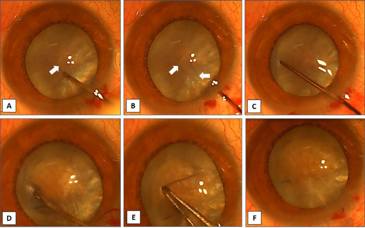
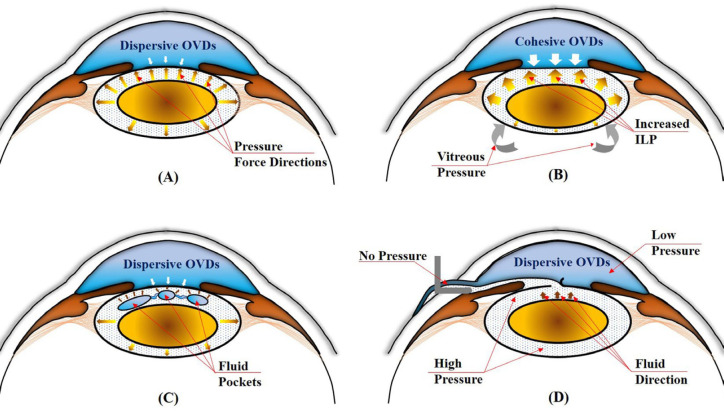
Methods: This prospective interventional study included 33 eyes with mature cataracts and evidence of subcapsular fluid spaces by slit lamp examination. For each patient, 20% mannitol was administered intravenously according to the bodyweight 1 h preoperatively. Under peribulbar anesthesia, a 2.2-mm main incision was made, and the anterior chamber was filled with a dispersive ophthalmic viscosurgical device. Using a bent-tip cystotome, a 2-mm curved incision was made in the center of the anterior capsule, which released subcortical fluid and was drained through compression of the posterior lip of the main incision using a spatula. Then, fine gentle milking in all quadrants around the puncture on the anterior lens capsule from the periphery toward the site of puncture using the blunt-edged spatula further assists drainage of subcortical fluid and breaks fine septa inside the lens to remove fluid from intralenticular fluid pocket collections.
Results: The study included 15 (45.5%) men and 18 (54.5%) women with a mean (standard deviation [SD]) of age of 63.2 (5.33) and 64.4 (6.21) years, respectively. The modified capsulorhexis technique was performed for 33 intumescent cataracts. Capsulorhexis was completed in all cases; capsulorhexis was easy in 31 (94%) eyes and difficult in 2 (6%) eyes. In the two difficult cases, radial extension occurred in one eye, and it was retrieved using the Little technique; the other case with radial tear was completed successfully using a retinal micro scissor from the other edge of the capsulorhexis until reaching an oval, continuous capsulorhexis.
Conclusions: This modified capsulorhexis technique with compression on the posterior lip of the main incision and capsule milking allowed for a safe, continuous curvilinear capsulorhexis. Further comparative studies are necessary to confirm our preliminary results.


 分享
分享
 求助内容:
求助内容: 应助结果提醒方式:
 扫码关注我们
