{"title":"Macular Imaging Characteristics in Children with Myelinated Retinal Nerve Fiber and High Myopia Syndrome.","authors":"Almila Sarıgül Sezenöz, Sibel Oto, İmren Akkoyun, Sezin Akça Bayar, Gürsel Yılmaz, Meriç Yavuz Çolak","doi":"10.4274/tjo.galenos.2023.27612","DOIUrl":null,"url":null,"abstract":"<p><strong>Objectives: </strong>To investigate the macular imaging features in patients with unilateral myelinated retinal nerve fiber (MRNF) and high myopia syndrome.</p><p><strong>Materials and methods: </strong>Six patients with unilateral MRNF and high myopia syndrome and 13 myopic controls were enrolled in this study. Spectral domain (SD) optical coherence tomography (OCT), SD enhanced depth imaging OCT, and OCT angiography (OCTA) imaging results of MRNF-affected eyes were compared with the fellow eyes and myopic controls.</p><p><strong>Results: </strong>All patients had abnormal foveal reflex and/or ectopia. No significant difference in retinal thickness parameters were noted between the groups. In OCT scans, posterior vitreous detachment (PVD) was observed in 4 out of the 6 MRNF-affected eyes. Regarding OCTA parameters, only a significant increase in acircularity index was noted in myelinated eyes (p=0.01).</p><p><strong>Conclusion: </strong>All patients demonstrated normal foveal contours, macular structure, and OCTA features except for a higher acircularity index. The incidence of PVD was notably increased in the myelinated eyes.</p>","PeriodicalId":23373,"journal":{"name":"Turkish Journal of Ophthalmology","volume":"53 4","pages":"234-240"},"PeriodicalIF":0.0000,"publicationDate":"2023-08-19","publicationTypes":"Journal Article","fieldsOfStudy":null,"isOpenAccess":false,"openAccessPdf":"https://ftp.ncbi.nlm.nih.gov/pub/pmc/oa_pdf/58/f1/TJO-53-234.PMC10442755.pdf","citationCount":"0","resultStr":null,"platform":"Semanticscholar","paperid":null,"PeriodicalName":"Turkish Journal of Ophthalmology","FirstCategoryId":"1085","ListUrlMain":"https://doi.org/10.4274/tjo.galenos.2023.27612","RegionNum":0,"RegionCategory":null,"ArticlePicture":[],"TitleCN":null,"AbstractTextCN":null,"PMCID":null,"EPubDate":"","PubModel":"","JCR":"Q3","JCRName":"Medicine","Score":null,"Total":0}
引用次数: 0
Abstract
Objectives: To investigate the macular imaging features in patients with unilateral myelinated retinal nerve fiber (MRNF) and high myopia syndrome.
Materials and methods: Six patients with unilateral MRNF and high myopia syndrome and 13 myopic controls were enrolled in this study. Spectral domain (SD) optical coherence tomography (OCT), SD enhanced depth imaging OCT, and OCT angiography (OCTA) imaging results of MRNF-affected eyes were compared with the fellow eyes and myopic controls.
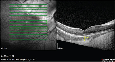
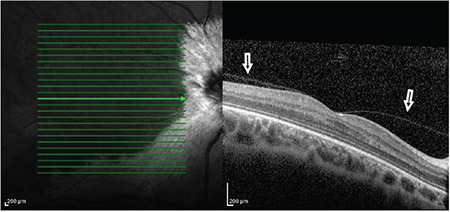
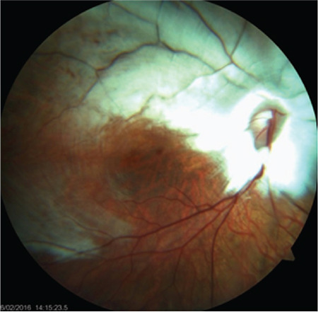
Results: All patients had abnormal foveal reflex and/or ectopia. No significant difference in retinal thickness parameters were noted between the groups. In OCT scans, posterior vitreous detachment (PVD) was observed in 4 out of the 6 MRNF-affected eyes. Regarding OCTA parameters, only a significant increase in acircularity index was noted in myelinated eyes (p=0.01).
Conclusion: All patients demonstrated normal foveal contours, macular structure, and OCTA features except for a higher acircularity index. The incidence of PVD was notably increased in the myelinated eyes.
期刊介绍:
The Turkish Journal of Ophthalmology (TJO) is the only scientific periodical publication of the Turkish Ophthalmological Association and has been published since January 1929. In its early years, the journal was published in Turkish and French. Although there were temporary interruptions in the publication of the journal due to various challenges, the Turkish Journal of Ophthalmology has been published continually from 1971 to the present. The target audience includes specialists and physicians in training in ophthalmology in all relevant disciplines.


 分享
分享
 求助内容:
求助内容: 应助结果提醒方式:
 扫码关注我们
