{"title":"Electron Microscopic Study in the Rat Model of Electrically Injured Myelopathy: Preliminary Report.","authors":"Je Hoon Jeong, Cheong Hoon Seo, Dae Hoon Lee","doi":"10.13004/kjnt.2023.19.e15","DOIUrl":null,"url":null,"abstract":"<p><strong>Objective: </strong>The patient with electrically injured myelopathy showed mild motor weakness without somatosensory pathway abnormalities. Few reports have been reported on the pathophysiological mechanisms of electrically injured myelopathy, and there is controversy about the exact pathological causes. This study aimed to investigate the ultrastructural changes in the electron microscopic findings of electrical spinal cord injury.</p><p><strong>Methods: </strong>Nine rats were used in this study. We performed 7 electrical shocks (frequency, 120 Hz; pulse width, 0.9 ms; duration, 3 seconds; current, 99 mA) using an electroconvulsive therapy (ECT) apparatus (57800 ECT unit; UGO BASILE). We used one ear and one contralateral hind limb as entry and exit sites, respectively. We only enrolled rats with hind limb weakness and performed electron microscopy evaluations of the spinal cord on the first day and 4 weeks after injury.</p><p><strong>Results: </strong>On the first day after injury, an electron microscopic examination showed a directly damaged area that appeared to be torn as physical damage, damaged myelin sheath, vacuolated axons in the myelin sheath, swollen Golgi apparatus, and injured mitochondria. Looking at changes in motor and sensory nerves, the sensory neurons showed recovered mitochondria and Golgi apparatus 4 weeks after injury; however, motor neurons still showed injured mitochondria, swollen Golgi apparatus, and endoplasmic reticulum.</p><p><strong>Conclusion: </strong>This study showed that recovery from ultrastructural injury was more rapid in sensory neurons than in motor neurons.</p>","PeriodicalId":36879,"journal":{"name":"Korean Journal of Neurotrauma","volume":"19 2","pages":"218-226"},"PeriodicalIF":0.0000,"publicationDate":"2023-06-01","publicationTypes":"Journal Article","fieldsOfStudy":null,"isOpenAccess":false,"openAccessPdf":"https://ftp.ncbi.nlm.nih.gov/pub/pmc/oa_pdf/39/55/kjn-19-218.PMC10329894.pdf","citationCount":"0","resultStr":null,"platform":"Semanticscholar","paperid":null,"PeriodicalName":"Korean Journal of Neurotrauma","FirstCategoryId":"1085","ListUrlMain":"https://doi.org/10.13004/kjnt.2023.19.e15","RegionNum":0,"RegionCategory":null,"ArticlePicture":[],"TitleCN":null,"AbstractTextCN":null,"PMCID":null,"EPubDate":"","PubModel":"","JCR":"Q3","JCRName":"Medicine","Score":null,"Total":0}
引用次数: 0
Abstract
Objective: The patient with electrically injured myelopathy showed mild motor weakness without somatosensory pathway abnormalities. Few reports have been reported on the pathophysiological mechanisms of electrically injured myelopathy, and there is controversy about the exact pathological causes. This study aimed to investigate the ultrastructural changes in the electron microscopic findings of electrical spinal cord injury.
Methods: Nine rats were used in this study. We performed 7 electrical shocks (frequency, 120 Hz; pulse width, 0.9 ms; duration, 3 seconds; current, 99 mA) using an electroconvulsive therapy (ECT) apparatus (57800 ECT unit; UGO BASILE). We used one ear and one contralateral hind limb as entry and exit sites, respectively. We only enrolled rats with hind limb weakness and performed electron microscopy evaluations of the spinal cord on the first day and 4 weeks after injury.
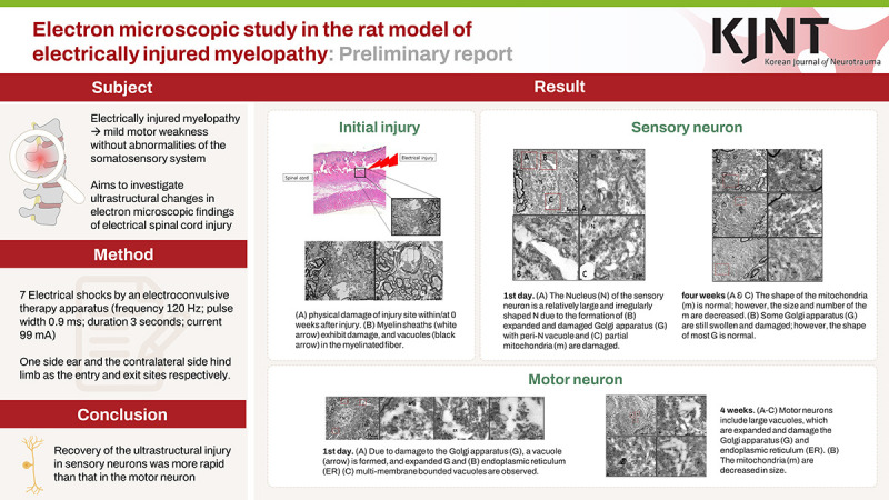
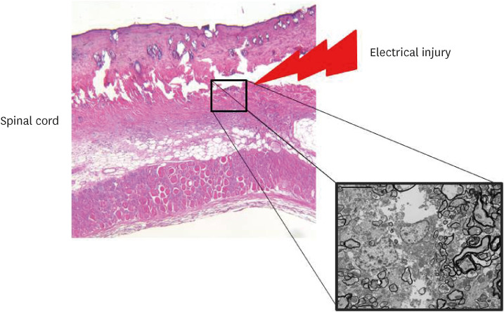
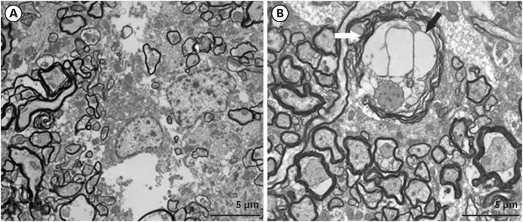
Results: On the first day after injury, an electron microscopic examination showed a directly damaged area that appeared to be torn as physical damage, damaged myelin sheath, vacuolated axons in the myelin sheath, swollen Golgi apparatus, and injured mitochondria. Looking at changes in motor and sensory nerves, the sensory neurons showed recovered mitochondria and Golgi apparatus 4 weeks after injury; however, motor neurons still showed injured mitochondria, swollen Golgi apparatus, and endoplasmic reticulum.
Conclusion: This study showed that recovery from ultrastructural injury was more rapid in sensory neurons than in motor neurons.


 分享
分享
 求助内容:
求助内容: 应助结果提醒方式:
 扫码关注我们
