Min Sun Bae, Hyo Young Kim, Hyunseung Oh, Bo Kyoung Seo
{"title":"Clinical applications of shear wave dispersion imaging for breast lesions: a pictorial essay.","authors":"Min Sun Bae, Hyo Young Kim, Hyunseung Oh, Bo Kyoung Seo","doi":"10.14366/usg.23079","DOIUrl":null,"url":null,"abstract":"<p><p>Shear wave dispersion (SWD) imaging is a newly developed ultrasound technology designed to evaluate the dispersion slope of shear waves, which is related to tissue viscosity. This advanced imaging technique holds potential for distinguishing malignant lesions from benign lesions and normal breast tissue. The SWD slope, as determined by shear wave elastography (SWE), could offer crucial insights into the characterization of breast lesions. This article presents SWE and SWD images of both malignant and benign breast lesions, in addition to normal breast tissue.</p>","PeriodicalId":54227,"journal":{"name":"Ultrasonography","volume":" ","pages":"589-599"},"PeriodicalIF":2.5000,"publicationDate":"2023-10-01","publicationTypes":"Journal Article","fieldsOfStudy":null,"isOpenAccess":false,"openAccessPdf":"https://ftp.ncbi.nlm.nih.gov/pub/pmc/oa_pdf/23/35/usg-23079.PMC10555686.pdf","citationCount":"0","resultStr":null,"platform":"Semanticscholar","paperid":null,"PeriodicalName":"Ultrasonography","FirstCategoryId":"3","ListUrlMain":"https://doi.org/10.14366/usg.23079","RegionNum":3,"RegionCategory":"医学","ArticlePicture":[],"TitleCN":null,"AbstractTextCN":null,"PMCID":null,"EPubDate":"2023/8/7 0:00:00","PubModel":"Epub","JCR":"Q2","JCRName":"RADIOLOGY, NUCLEAR MEDICINE & MEDICAL IMAGING","Score":null,"Total":0}
引用次数: 0
Abstract
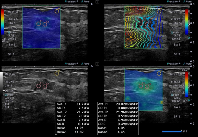
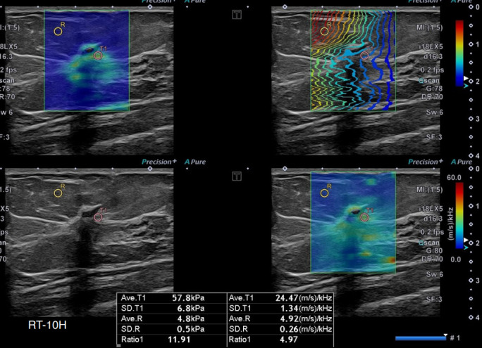
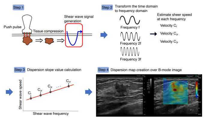
Shear wave dispersion (SWD) imaging is a newly developed ultrasound technology designed to evaluate the dispersion slope of shear waves, which is related to tissue viscosity. This advanced imaging technique holds potential for distinguishing malignant lesions from benign lesions and normal breast tissue. The SWD slope, as determined by shear wave elastography (SWE), could offer crucial insights into the characterization of breast lesions. This article presents SWE and SWD images of both malignant and benign breast lesions, in addition to normal breast tissue.
UltrasonographyMedicine-Radiology, Nuclear Medicine and Imaging
CiteScore
5.10
自引率
6.50%
发文量
78
审稿时长
15 weeks
期刊介绍:
Ultrasonography, the official English-language journal of the Korean Society of Ultrasound in Medicine (KSUM), is an international peer-reviewed academic journal dedicated to practice, research, technology, and education dealing with medical ultrasound. It is renamed from the Journal of Korean Society of Ultrasound in Medicine in January 2014, and published four times per year: January 1, April 1, July 1, and October 1. Original articles, technical notes, topical reviews, perspectives, pictorial essays, and timely editorial materials are published in Ultrasonography covering state-of-the-art content.
Ultrasonography aims to provide updated information on new diagnostic concepts and technical developments, including experimental animal studies using new equipment in addition to well-designed reviews of contemporary issues in patient care. Along with running KSUM Open, the annual international congress of KSUM, Ultrasonography also serves as a medium for cooperation among physicians and specialists from around the world who are focusing on various ultrasound technology and disease problems and relevant basic science.


 分享
分享
 求助内容:
求助内容: 应助结果提醒方式:
 扫码关注我们
