Jonathan Noël, Anya Mascarenhas, Chibueze A Nwaiwu, Yao Liu, Marcio Moschovas, Vasiliy E Buharin, John Oberlin, Saloni Mehrotra, Alyson F Dechert, Peter C W Kim, Vipul Patel
{"title":"Laser speckle contrast imaging compared with indocyanine green in renal perfusion of a porcine model.","authors":"Jonathan Noël, Anya Mascarenhas, Chibueze A Nwaiwu, Yao Liu, Marcio Moschovas, Vasiliy E Buharin, John Oberlin, Saloni Mehrotra, Alyson F Dechert, Peter C W Kim, Vipul Patel","doi":"10.1097/CU9.0000000000000155","DOIUrl":null,"url":null,"abstract":"<p><strong>Background: </strong>When viewed under near-infrared light, indocyanine green (ICG) signal for kidney perfusion can be utilized in partial nephrectomy. Laser speckle contrast imaging (LSCI) uses coherent light to detect perfusion during real-time laparoscopic surgery.</p><p><strong>Materials and methods: </strong>Laser speckle contrast imaging or ActivSight, an imaging sensor adapter, was used during laparoscopy of an anesthetized porcine kidney model. ActivSight's \"perfusion mode\" and \"quantification mode\" displayed the blood flow as a heatmap and numerical signal intensity, respectively.</p><p><strong>Results: </strong>After the upper segmental renal artery was clamped, ICG was seen in the lower pole, and LSCI showed low unit (dark color) quantification and perfusion in the upper pole. Indocyanine green was retained in the lower pole after the upper segmental artery was unclamped, and LSCI perfusion was demonstrated in the entire kidney.</p><p><strong>Conclusions: </strong>Laser speckle contrast imaging is a dye-free, repeatable, real-time adjunct for renal parenchymal perfusion assessment applicable to minimally invasive renal surgery to complement the technology of ICG near-infrared fluorescence and advance digital surgery.</p>","PeriodicalId":39147,"journal":{"name":"Current Urology","volume":"17 2","pages":"141-145"},"PeriodicalIF":1.3000,"publicationDate":"2023-06-01","publicationTypes":"Journal Article","fieldsOfStudy":null,"isOpenAccess":false,"openAccessPdf":"https://ftp.ncbi.nlm.nih.gov/pub/pmc/oa_pdf/8f/f2/curr-urol-17-141.PMC10489255.pdf","citationCount":"0","resultStr":null,"platform":"Semanticscholar","paperid":null,"PeriodicalName":"Current Urology","FirstCategoryId":"1085","ListUrlMain":"https://doi.org/10.1097/CU9.0000000000000155","RegionNum":4,"RegionCategory":"医学","ArticlePicture":[],"TitleCN":null,"AbstractTextCN":null,"PMCID":null,"EPubDate":"","PubModel":"","JCR":"Q4","JCRName":"UROLOGY & NEPHROLOGY","Score":null,"Total":0}
引用次数: 0
Abstract
Background: When viewed under near-infrared light, indocyanine green (ICG) signal for kidney perfusion can be utilized in partial nephrectomy. Laser speckle contrast imaging (LSCI) uses coherent light to detect perfusion during real-time laparoscopic surgery.
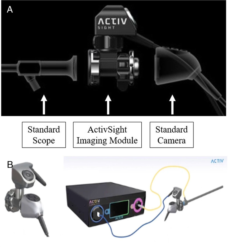
Materials and methods: Laser speckle contrast imaging or ActivSight, an imaging sensor adapter, was used during laparoscopy of an anesthetized porcine kidney model. ActivSight's "perfusion mode" and "quantification mode" displayed the blood flow as a heatmap and numerical signal intensity, respectively.
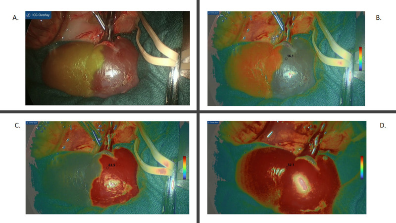
Results: After the upper segmental renal artery was clamped, ICG was seen in the lower pole, and LSCI showed low unit (dark color) quantification and perfusion in the upper pole. Indocyanine green was retained in the lower pole after the upper segmental artery was unclamped, and LSCI perfusion was demonstrated in the entire kidney.
Conclusions: Laser speckle contrast imaging is a dye-free, repeatable, real-time adjunct for renal parenchymal perfusion assessment applicable to minimally invasive renal surgery to complement the technology of ICG near-infrared fluorescence and advance digital surgery.


 分享
分享
 求助内容:
求助内容: 应助结果提醒方式:
 扫码关注我们
