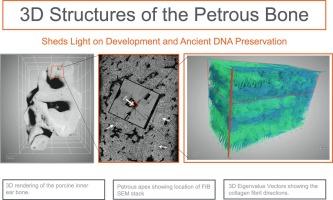
We report on the 3D ultrastructure of the mineralized petrous bone of mature pig using focused ion beam – scanning electron microscopy (FIB-SEM). We divide the petrous bone into two zones based on the degree of mineralization; one zone close to the otic chamber has higher mineral density than the second zone further away from the otic chamber. The hypermineralization of the petrous bone results in the collagen D-banding being poorly revealed in the lower mineral density zone (LMD), and absent in the high mineral density zone (HMD). We therefore could not use D-banding to decipher the 3D structure of the collagen assembly. Instead we exploited the anisotropy option in the Dragonfly image processing software to visualize the less mineralized collagen fibrils and/or nanopores that surround the more mineralized zones known as tesselles. This approach therefore indirectly tracks the orientations of the collagen fibrils in the matrix itself. We show that the HMD bone has a structure similar to that of woven bone, and the LMD is composed of lamellar bone with a plywood-like structural motif. This agrees with the fact that the bone close to the otic chamber is fetal bone and is not remodeled. The lamellar structure of the bone further away from the otic chamber is consistent with modeling/remodeling. The absence of the less mineralized collagen fibrils and nanopores resulting from the confluence of the mineral tesselles may contribute to shielding DNA during diagenesis. We show that anisotropy evaluation of the less mineralized collagen fibrils could be a useful tool to analyze bone ultrastructures and in particular the directionality of collagen fibril bundles that make up the bone matrix.


 分享
分享
 求助内容:
求助内容: 应助结果提醒方式:
 扫码关注我们
