Cristina Cardenal Peralta , Paul Vandroux , Lea Neumann-Arnold , Michel Panvert , Jérôme Fagart , Wolfgang Seufert , Yves Mechulam , Emmanuelle Schmitt
{"title":"Binding of human Cdc123 to eIF2γ","authors":"Cristina Cardenal Peralta , Paul Vandroux , Lea Neumann-Arnold , Michel Panvert , Jérôme Fagart , Wolfgang Seufert , Yves Mechulam , Emmanuelle Schmitt","doi":"10.1016/j.jsb.2023.108006","DOIUrl":null,"url":null,"abstract":"<div><p>Eukaryotic initiation factor 2 (eIF2) plays a key role in protein synthesis and in its regulation. The assembly of this heterotrimeric factor is facilitated by Cdc123, a member of the ATP grasp family that binds the γ subunit of eIF2. Notably, some mutations related to MEHMO syndrome, an X-linked intellectual disability, affect Cdc123-mediated eIF2 assembly. The mechanism of action of Cdc123 is unclear and structural information for the human protein is awaited. Here, the crystallographic structure of human Cdc123 (Hs-Cdc123) bound to domain 3 of human eIF2γ (Hs-eIF2γD3) was determined. The structure shows that the domain 3 of eIF2γ is bound to domain 1 of Cdc123. In addition, the long C-terminal region of Hs-Cdc123 provides a link between the ATP and Hs-eIF2γD3 binding sites. A thermal shift assay shows that ATP is tightly bound to Cdc123 whereas the affinity of ADP is much smaller. Yeast cell viability experiments, western blot analysis and two-hybrid assays show that ATP is important for the function of Hs-Cdc123 in eIF2 assembly. These data and recent findings allow us to propose a refined model to explain the mechanism of action of Cdc123 in eIF2 assembly.</p></div>","PeriodicalId":17074,"journal":{"name":"Journal of structural biology","volume":"215 3","pages":"Article 108006"},"PeriodicalIF":2.7000,"publicationDate":"2023-09-01","publicationTypes":"Journal Article","fieldsOfStudy":null,"isOpenAccess":false,"openAccessPdf":"","citationCount":"0","resultStr":null,"platform":"Semanticscholar","paperid":null,"PeriodicalName":"Journal of structural biology","FirstCategoryId":"99","ListUrlMain":"https://www.sciencedirect.com/science/article/pii/S1047847723000692","RegionNum":3,"RegionCategory":"生物学","ArticlePicture":[],"TitleCN":null,"AbstractTextCN":null,"PMCID":null,"EPubDate":"2023/7/27 0:00:00","PubModel":"Epub","JCR":"Q3","JCRName":"BIOCHEMISTRY & MOLECULAR BIOLOGY","Score":null,"Total":0}
引用次数: 0
Abstract
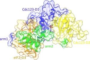
Eukaryotic initiation factor 2 (eIF2) plays a key role in protein synthesis and in its regulation. The assembly of this heterotrimeric factor is facilitated by Cdc123, a member of the ATP grasp family that binds the γ subunit of eIF2. Notably, some mutations related to MEHMO syndrome, an X-linked intellectual disability, affect Cdc123-mediated eIF2 assembly. The mechanism of action of Cdc123 is unclear and structural information for the human protein is awaited. Here, the crystallographic structure of human Cdc123 (Hs-Cdc123) bound to domain 3 of human eIF2γ (Hs-eIF2γD3) was determined. The structure shows that the domain 3 of eIF2γ is bound to domain 1 of Cdc123. In addition, the long C-terminal region of Hs-Cdc123 provides a link between the ATP and Hs-eIF2γD3 binding sites. A thermal shift assay shows that ATP is tightly bound to Cdc123 whereas the affinity of ADP is much smaller. Yeast cell viability experiments, western blot analysis and two-hybrid assays show that ATP is important for the function of Hs-Cdc123 in eIF2 assembly. These data and recent findings allow us to propose a refined model to explain the mechanism of action of Cdc123 in eIF2 assembly.
期刊介绍:
Journal of Structural Biology (JSB) has an open access mirror journal, the Journal of Structural Biology: X (JSBX), sharing the same aims and scope, editorial team, submission system and rigorous peer review. Since both journals share the same editorial system, you may submit your manuscript via either journal homepage. You will be prompted during submission (and revision) to choose in which to publish your article. The editors and reviewers are not aware of the choice you made until the article has been published online. JSB and JSBX publish papers dealing with the structural analysis of living material at every level of organization by all methods that lead to an understanding of biological function in terms of molecular and supermolecular structure.
Techniques covered include:
• Light microscopy including confocal microscopy
• All types of electron microscopy
• X-ray diffraction
• Nuclear magnetic resonance
• Scanning force microscopy, scanning probe microscopy, and tunneling microscopy
• Digital image processing
• Computational insights into structure


 分享
分享
 求助内容:
求助内容: 应助结果提醒方式:
 扫码关注我们
