Yogan Kisten, Laurent Arnaud, Adrian Levitsky, Noémi Györi, Per Larsson, Aase Hensvold, Anca Catrina, Erik Af Klint, Hamed Rezaei
{"title":"Distinct Fluorescence Optical Imaging Patient Clusters Emerge for Seropositive and Seronegative Rheumatoid Arthritis.","authors":"Yogan Kisten, Laurent Arnaud, Adrian Levitsky, Noémi Györi, Per Larsson, Aase Hensvold, Anca Catrina, Erik Af Klint, Hamed Rezaei","doi":"10.1002/acr2.11599","DOIUrl":null,"url":null,"abstract":"<p><strong>Objective: </strong>To investigate whether digital activity fluorescence optical imaging (FOI) patterns of inflammation can identify distinct rheumatoid arthritis (RA) phenotypes.</p><p><strong>Methods: </strong>The hands of newly diagnosed patients with RA were evaluated by clinical examination, musculoskeletal ultrasound, and FOI. Inflammation on FOI was defined when capillary leakage and/or fluorophore perfusion was present. The FOI composite image was quantified into a digital disease activity (DACT) score, using novel computerized algorithms. Unsupervised clustering on FOI inflammatory patterns was used to identify subgroups of patients relative to anticyclic citrullinated peptides (ACPA) and/or rheumatoid factor (RF).</p><p><strong>Results: </strong>Of 1326 examined hand joints in 39 patients with RA (72% female; 56% ever-smokers; 54% RF positive and 69% ACPA positive), 400 (30%) showed inflammation by FOI, and 95% (37 of 39) of patients had DACT-FOI scores greater than 1. Unsupervised analysis on FOI patterns revealed two patient clusters, cluster 1 (n = 29) and cluster 2 (n = 10). The proportion of seropositive patients was significantly higher in cluster 1 versus cluster 2 (90%, 26 of 29 vs. 30%, 3 of 10; P < 0.01), whereas C-reactive-protein levels (minimum-maximum) were significantly higher in cluster 2 (20 mg/l [1-102]) versus cluster 1 (2 mg/l [0-119]; P = 0.01). A wider variety and proportion of inflamed joints emerged for patients with RA in cluster 2 versus cluster 1, in which inflammation was more concentrated around the wrists and the right metacarpophalangeal 2 (MCP2), bilateral MCP3, and, to a lesser degree, left MCP2 and proximal interphalangeal joint and tendon regions. Cluster 1 displayed lower mean (±SD) DACT scores compared with cluster 2 (3.6 ± 2.1 vs. 5.4 ± 2.1; P = 0.03).</p><p><strong>Conclusion: </strong>FOI-based digital quantification of hand joint inflammation revealed two distinct RA subpopulations with and without ACPA and RF related autoantibodies.</p>","PeriodicalId":7084,"journal":{"name":"ACR Open Rheumatology","volume":"5 9","pages":"474-480"},"PeriodicalIF":0.0000,"publicationDate":"2023-09-01","publicationTypes":"Journal Article","fieldsOfStudy":null,"isOpenAccess":false,"openAccessPdf":"https://ftp.ncbi.nlm.nih.gov/pub/pmc/oa_pdf/76/94/ACR2-5-474.PMC10502810.pdf","citationCount":"0","resultStr":null,"platform":"Semanticscholar","paperid":null,"PeriodicalName":"ACR Open Rheumatology","FirstCategoryId":"1085","ListUrlMain":"https://doi.org/10.1002/acr2.11599","RegionNum":0,"RegionCategory":null,"ArticlePicture":[],"TitleCN":null,"AbstractTextCN":null,"PMCID":null,"EPubDate":"","PubModel":"","JCR":"","JCRName":"","Score":null,"Total":0}
引用次数: 0
Abstract
Objective: To investigate whether digital activity fluorescence optical imaging (FOI) patterns of inflammation can identify distinct rheumatoid arthritis (RA) phenotypes.
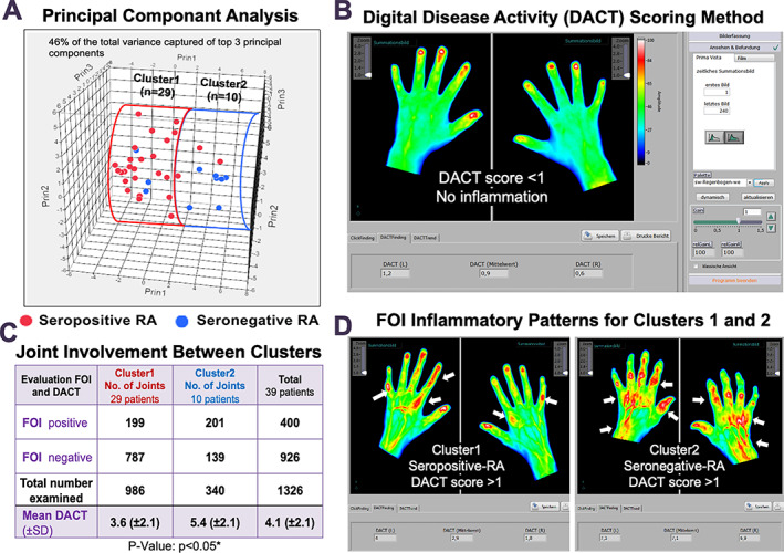
Methods: The hands of newly diagnosed patients with RA were evaluated by clinical examination, musculoskeletal ultrasound, and FOI. Inflammation on FOI was defined when capillary leakage and/or fluorophore perfusion was present. The FOI composite image was quantified into a digital disease activity (DACT) score, using novel computerized algorithms. Unsupervised clustering on FOI inflammatory patterns was used to identify subgroups of patients relative to anticyclic citrullinated peptides (ACPA) and/or rheumatoid factor (RF).
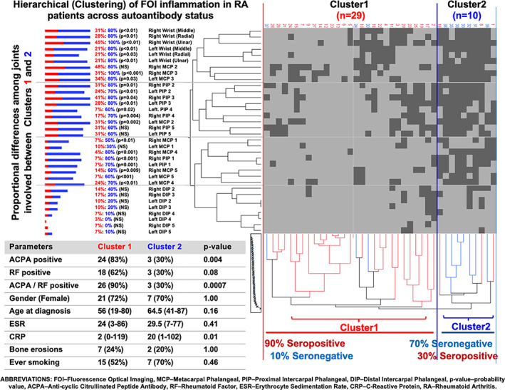
Results: Of 1326 examined hand joints in 39 patients with RA (72% female; 56% ever-smokers; 54% RF positive and 69% ACPA positive), 400 (30%) showed inflammation by FOI, and 95% (37 of 39) of patients had DACT-FOI scores greater than 1. Unsupervised analysis on FOI patterns revealed two patient clusters, cluster 1 (n = 29) and cluster 2 (n = 10). The proportion of seropositive patients was significantly higher in cluster 1 versus cluster 2 (90%, 26 of 29 vs. 30%, 3 of 10; P < 0.01), whereas C-reactive-protein levels (minimum-maximum) were significantly higher in cluster 2 (20 mg/l [1-102]) versus cluster 1 (2 mg/l [0-119]; P = 0.01). A wider variety and proportion of inflamed joints emerged for patients with RA in cluster 2 versus cluster 1, in which inflammation was more concentrated around the wrists and the right metacarpophalangeal 2 (MCP2), bilateral MCP3, and, to a lesser degree, left MCP2 and proximal interphalangeal joint and tendon regions. Cluster 1 displayed lower mean (±SD) DACT scores compared with cluster 2 (3.6 ± 2.1 vs. 5.4 ± 2.1; P = 0.03).
Conclusion: FOI-based digital quantification of hand joint inflammation revealed two distinct RA subpopulations with and without ACPA and RF related autoantibodies.


 分享
分享

 求助内容:
求助内容: 应助结果提醒方式:
 扫码关注我们
