Relation between Condyle Horizontal Angle and Intercondylar Angle with Disc Displacement in Patients with Temporomandibular Joint Disorders: An MRI Evaluation.
{"title":"Relation between Condyle Horizontal Angle and Intercondylar Angle with Disc Displacement in Patients with Temporomandibular Joint Disorders: An MRI Evaluation.","authors":"Shahriar Shahab, Zahra Amoozad Khalili, Elham Emami Meybodi, Morteza Banakar","doi":"10.1155/2023/3846525","DOIUrl":null,"url":null,"abstract":"<p><strong>Background: </strong>Internal derangement (ID) is the most common cause of temporomandibular disorders (TMDs) and extensively affects the articular disc function. The anterior disc displacement is among the most important findings in ID. Knowledge about the etiology of this condition is imperative, and the role of structural parameters in the development of TMDs has not been well evaluated.</p><p><strong>Objectives: </strong>This study aimed to assess the relationship between condylar angulation and intercondylar angle with anterior disc displacement in patients with TMD using magnetic resonance imaging (MRI).</p><p><strong>Materials and methods: </strong>This case-control study evaluated 31 temporomandibular joints with internal derangement and 57 normal joints. The data retrieved from MRI included disc position in the open mouth (normal, anterior disc displacement with a reduction (DDWR) and without reduction (DDWOR), and posterior displacement (PD)), horizontal condylar angle categorized as normal (10 to 30° angle) and abnormal (<10° and >30°), and intercondylar angle. Chi-square test, <i>T</i>-test, and Fisher's exact were done to assess the relationship between horizontal condylar angle and intercondylar angle in patients with TMDs with DDWR and DDWOR compared with the control group.</p><p><strong>Results: </strong>Patients with DDWR and DDWOR had higher odds of abnormal horizontal condylar angle, particularly >30° angle, which was a significant correlation (odds ratio of 0.19 and 8.3, respectively). The intercondylar angle in the patients with disc displacement was significantly smaller compared to the control group.</p><p><strong>Conclusion: </strong>Disc displacement was correlated with abnormal horizontal angle (particularly < 30) and smaller intercondylar angle compared with the control group.</p>","PeriodicalId":51864,"journal":{"name":"Radiology Research and Practice","volume":"2023 ","pages":"3846525"},"PeriodicalIF":1.5000,"publicationDate":"2023-09-08","publicationTypes":"Journal Article","fieldsOfStudy":null,"isOpenAccess":false,"openAccessPdf":"https://www.ncbi.nlm.nih.gov/pmc/articles/PMC10504042/pdf/","citationCount":"0","resultStr":null,"platform":"Semanticscholar","paperid":null,"PeriodicalName":"Radiology Research and Practice","FirstCategoryId":"1085","ListUrlMain":"https://doi.org/10.1155/2023/3846525","RegionNum":0,"RegionCategory":null,"ArticlePicture":[],"TitleCN":null,"AbstractTextCN":null,"PMCID":null,"EPubDate":"2023/1/1 0:00:00","PubModel":"eCollection","JCR":"Q2","JCRName":"RADIOLOGY, NUCLEAR MEDICINE & MEDICAL IMAGING","Score":null,"Total":0}
引用次数: 0
Abstract
Background: Internal derangement (ID) is the most common cause of temporomandibular disorders (TMDs) and extensively affects the articular disc function. The anterior disc displacement is among the most important findings in ID. Knowledge about the etiology of this condition is imperative, and the role of structural parameters in the development of TMDs has not been well evaluated.
Objectives: This study aimed to assess the relationship between condylar angulation and intercondylar angle with anterior disc displacement in patients with TMD using magnetic resonance imaging (MRI).
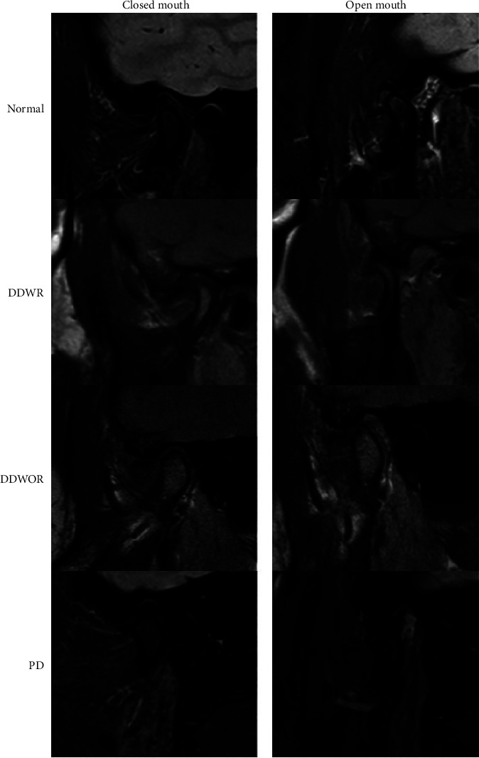
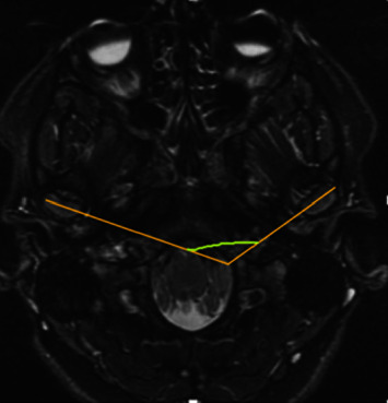
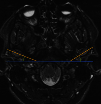
Materials and methods: This case-control study evaluated 31 temporomandibular joints with internal derangement and 57 normal joints. The data retrieved from MRI included disc position in the open mouth (normal, anterior disc displacement with a reduction (DDWR) and without reduction (DDWOR), and posterior displacement (PD)), horizontal condylar angle categorized as normal (10 to 30° angle) and abnormal (<10° and >30°), and intercondylar angle. Chi-square test, T-test, and Fisher's exact were done to assess the relationship between horizontal condylar angle and intercondylar angle in patients with TMDs with DDWR and DDWOR compared with the control group.
Results: Patients with DDWR and DDWOR had higher odds of abnormal horizontal condylar angle, particularly >30° angle, which was a significant correlation (odds ratio of 0.19 and 8.3, respectively). The intercondylar angle in the patients with disc displacement was significantly smaller compared to the control group.
Conclusion: Disc displacement was correlated with abnormal horizontal angle (particularly < 30) and smaller intercondylar angle compared with the control group.
期刊介绍:
Radiology Research and Practice is a peer-reviewed, Open Access journal that publishes articles on all areas of medical imaging. The journal promotes evidence-based radiology practice though the publication of original research, reviews, and clinical studies for a multidisciplinary audience. Radiology Research and Practice is archived in Portico, which provides permanent archiving for electronic scholarly journals, as well as via the LOCKSS initiative. It operates a fully open access publishing model which allows open global access to its published content. This model is supported through Article Processing Charges. For more information on Article Processing charges in gen


 分享
分享
 求助内容:
求助内容: 应助结果提醒方式:
 扫码关注我们
