Dariia Yosypyshyn, Domantė Kučikienė, Inez Ramakers, Jörg B Schulz, Kathrin Reetz, Ana Sofia Costa
{"title":"Clinical characteristics of patients with suspected Alzheimer's disease within a CSF Aß-ratio grey zone.","authors":"Dariia Yosypyshyn, Domantė Kučikienė, Inez Ramakers, Jörg B Schulz, Kathrin Reetz, Ana Sofia Costa","doi":"10.1186/s42466-023-00262-8","DOIUrl":null,"url":null,"abstract":"<p><strong>Background: </strong>The AT(N) research framework for Alzheimer's disease (AD) remains unclear on how to best deal with borderline cases. Our aim was to characterise patients with suspected AD with a borderline Aß<sub>1-42</sub>/Aß<sub>1-40</sub> ratio in cerebrospinal fluid.</p><p><strong>Methods: </strong>We analysed retrospective data from two cohorts (memory clinic cohort and ADNI) of patients (n = 63) with an Aß<sub>1-42</sub>/Aß<sub>1-40</sub> ratio within a predefined borderline area-Q<sub>1</sub> above the validated cut-off value(grey zone). We compared demographic, clinical, neuropsychological and neuroimaging features between grey zone patients and patients with low Aß<sub>1-42</sub> (normal Aß ratio but pathological Aß<sub>1-42</sub>, n = 42) and patients with AD (pathological Aß, P-Tau, und T-Tau, n = 80).</p><p><strong>Results: </strong>Patients had mild cognitive impairment or mild dementia and a median age of 72 years. Demographic and general clinical characteristics did not differ between the groups. Patients in the grey zone group were the least impaired in cognition. However, they overlapped with the low Aß<sub>1-42</sub> group in verbal episodic memory performance, especially in delayed recall and recognition. The grey zone group had less severe medial temporal atrophy, but mild posterior atrophy and mild white matter hyperintensities, similar to the low Aß<sub>1-42</sub> group.</p><p><strong>Conclusions: </strong>Patients in the Aß ratio grey zone were less impaired, but showed clinical overlap with patients on the AD continuum. These borderline patients may be at an earlier disease stage. Assuming an increased risk of AD and progressive cognitive decline, careful consideration of clinical follow-up is recommended when using dichotomous approaches to classify Aß status.</p>","PeriodicalId":19169,"journal":{"name":"Neurological Research and Practice","volume":"5 1","pages":"40"},"PeriodicalIF":0.0000,"publicationDate":"2023-08-03","publicationTypes":"Journal Article","fieldsOfStudy":null,"isOpenAccess":false,"openAccessPdf":"https://www.ncbi.nlm.nih.gov/pmc/articles/PMC10398972/pdf/","citationCount":"0","resultStr":null,"platform":"Semanticscholar","paperid":null,"PeriodicalName":"Neurological Research and Practice","FirstCategoryId":"1085","ListUrlMain":"https://doi.org/10.1186/s42466-023-00262-8","RegionNum":0,"RegionCategory":null,"ArticlePicture":[],"TitleCN":null,"AbstractTextCN":null,"PMCID":null,"EPubDate":"","PubModel":"","JCR":"","JCRName":"","Score":null,"Total":0}
引用次数: 0
Abstract
Background: The AT(N) research framework for Alzheimer's disease (AD) remains unclear on how to best deal with borderline cases. Our aim was to characterise patients with suspected AD with a borderline Aß1-42/Aß1-40 ratio in cerebrospinal fluid.
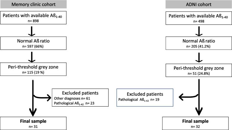
Methods: We analysed retrospective data from two cohorts (memory clinic cohort and ADNI) of patients (n = 63) with an Aß1-42/Aß1-40 ratio within a predefined borderline area-Q1 above the validated cut-off value(grey zone). We compared demographic, clinical, neuropsychological and neuroimaging features between grey zone patients and patients with low Aß1-42 (normal Aß ratio but pathological Aß1-42, n = 42) and patients with AD (pathological Aß, P-Tau, und T-Tau, n = 80).
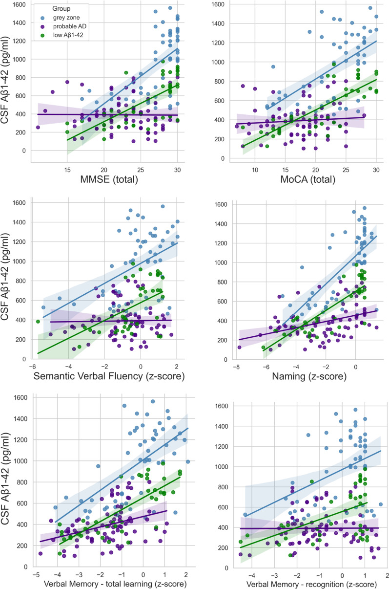
Results: Patients had mild cognitive impairment or mild dementia and a median age of 72 years. Demographic and general clinical characteristics did not differ between the groups. Patients in the grey zone group were the least impaired in cognition. However, they overlapped with the low Aß1-42 group in verbal episodic memory performance, especially in delayed recall and recognition. The grey zone group had less severe medial temporal atrophy, but mild posterior atrophy and mild white matter hyperintensities, similar to the low Aß1-42 group.
Conclusions: Patients in the Aß ratio grey zone were less impaired, but showed clinical overlap with patients on the AD continuum. These borderline patients may be at an earlier disease stage. Assuming an increased risk of AD and progressive cognitive decline, careful consideration of clinical follow-up is recommended when using dichotomous approaches to classify Aß status.
背景:对于阿尔茨海默病(AD)的AT(N)研究框架仍然不清楚如何最好地处理边缘性病例。我们的目的是确定脑脊液中a ß1-42/ a ß1-40比值为交界值的疑似AD患者的特征。方法:我们分析了来自两个队列(记忆诊所队列和ADNI)的患者(n = 63)的回顾性数据,这些患者的a ß1-42/ a ß1-40比率在预先确定的边界区域内-高于有效临界值(灰色地带)的q1。我们比较了灰色地带患者、低Aß1-42患者(正常Aß1-42但病理性Aß1-42, n = 42)和AD患者(病理性Aß、P-Tau和T-Tau, n = 80)的人口学、临床、神经心理学和神经影像学特征。结果:患者有轻度认知障碍或轻度痴呆,中位年龄72岁。人口学和一般临床特征在两组之间没有差异。灰色地带组患者的认知受损程度最小。然而,他们在言语情景记忆表现上与低Aß1-42组重叠,尤其是在延迟回忆和识别方面。灰色区组内侧颞叶萎缩较轻,但后部轻度萎缩,白质轻度高,与低Aß1-42组相似。结论:阿斯比灰色地带的患者受损程度较轻,但与AD连续体上的患者存在临床重叠。这些边缘患者可能处于疾病早期阶段。假设阿尔茨海默病的风险增加和认知能力的进行性下降,建议在使用二分类方法对asb状态进行分类时仔细考虑临床随访。


 分享
分享

 求助内容:
求助内容: 应助结果提醒方式:
 扫码关注我们
