{"title":"The \"<i>ghost sign</i>\": focal paleness as a novel marker of an inverted colonic diverticulum.","authors":"Vincent Zimmer","doi":"10.1097/j.pbj.0000000000000221","DOIUrl":null,"url":null,"abstract":"To the Editor: A 59-year-old male patient presented for surveillance colonoscopy on an outpatient basis. During scope advancement, besides a multitude of inconspicuous diverticula in the sigmoid with only occasional typical diverticula in the higher colon, a pale elevated lesion was noted behind a fold in the ascending colon (Fig. 1A). Further characterization on withdrawal indicated a normal mucosal appearance and a central umbilication on white light (Fig. 1B) and narrow-band imaging (Fig. 1C), likewise, however, highlighting the change in color in comparison with the background mucosa. To unequivocally demonstrate the nature of the lesion as an inverted diverticulum, we cautiously “deinverted” the diverticulum with the tip of a standard biopsy forceps (Fig. 1D). Albeit not per se mutually exclusive, clear-cut differentiation of polys from polypoid inverted colonic diverticula (ICD) as their typical endoscopic presentation is usually straightforward and essential to avoid undue endoscopic resection with inadvertent perforation, entailing potentially serious medical and medicolegal sequelae. Some endoscopic features, such as surrounding","PeriodicalId":74479,"journal":{"name":"Porto biomedical journal","volume":"8 4","pages":"e221"},"PeriodicalIF":0.0000,"publicationDate":"2023-07-01","publicationTypes":"Journal Article","fieldsOfStudy":null,"isOpenAccess":false,"openAccessPdf":"https://www.ncbi.nlm.nih.gov/pmc/articles/PMC10400048/pdf/","citationCount":"0","resultStr":null,"platform":"Semanticscholar","paperid":null,"PeriodicalName":"Porto biomedical journal","FirstCategoryId":"1085","ListUrlMain":"https://doi.org/10.1097/j.pbj.0000000000000221","RegionNum":0,"RegionCategory":null,"ArticlePicture":[],"TitleCN":null,"AbstractTextCN":null,"PMCID":null,"EPubDate":"","PubModel":"","JCR":"","JCRName":"","Score":null,"Total":0}
引用次数: 0
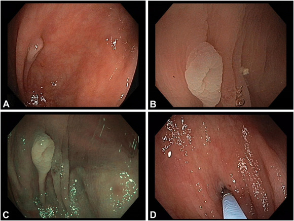
Abstract
To the Editor: A 59-year-old male patient presented for surveillance colonoscopy on an outpatient basis. During scope advancement, besides a multitude of inconspicuous diverticula in the sigmoid with only occasional typical diverticula in the higher colon, a pale elevated lesion was noted behind a fold in the ascending colon (Fig. 1A). Further characterization on withdrawal indicated a normal mucosal appearance and a central umbilication on white light (Fig. 1B) and narrow-band imaging (Fig. 1C), likewise, however, highlighting the change in color in comparison with the background mucosa. To unequivocally demonstrate the nature of the lesion as an inverted diverticulum, we cautiously “deinverted” the diverticulum with the tip of a standard biopsy forceps (Fig. 1D). Albeit not per se mutually exclusive, clear-cut differentiation of polys from polypoid inverted colonic diverticula (ICD) as their typical endoscopic presentation is usually straightforward and essential to avoid undue endoscopic resection with inadvertent perforation, entailing potentially serious medical and medicolegal sequelae. Some endoscopic features, such as surrounding


 分享
分享

 求助内容:
求助内容: 应助结果提醒方式:
 扫码关注我们
