{"title":"Herpes Proctitis in Men Mimicking Rectal Adenocarcinoma: Two Cases of an Easily Overlooked Diagnosis in the Proximal Rectum.","authors":"Jing Sun, Reenu Malhotra, Lakshmi Ananthakrishnan, Purva Gopal","doi":"10.1155/2023/6947960","DOIUrl":null,"url":null,"abstract":"<p><p>We describe two cases of rectal herpes simplex virus (HSV) infection in men that clinically mimicked rectal adenocarcinoma. Herpes infection in this location more commonly presents as an anal mass with viral inclusions in squamous epithelial cells. We report these cases to increase awareness of the unusual presentation as a proximal rectal mass with viral inclusions in endothelial cell nuclei. One patient was HIV-positive, and the other one had a history of having sex with men (MSM). Both patients had a thickened rectal wall with prominent lymphadenopathy on computed tomography (CT) scan, suspecting for malignancy. Biopsy showed abundant granulation tissue, necrosis, and inflammatory infiltrate composed predominantly of lymphocytes with admixed numerous plasma cells, eosinophils, and neutrophils. Rare granulation tissue vessels were lined by endothelial cells with nuclear molding and chromatin margination, and nuclei that were positive for HSV immunohistochemistry (IHC). One patient had confirmatory viral culture from biopsy of the ulcerated rectal mass. Both patients had symptom resolution following treatment for HSV. HSV should be considered in the differential diagnosis of rectal inflammatory masses, particularly in immunocompromised, HIV-positive, and MSM patients.</p>","PeriodicalId":45638,"journal":{"name":"Case Reports in Pathology","volume":"2023 ","pages":"6947960"},"PeriodicalIF":0.5000,"publicationDate":"2023-01-01","publicationTypes":"Journal Article","fieldsOfStudy":null,"isOpenAccess":false,"openAccessPdf":"https://www.ncbi.nlm.nih.gov/pmc/articles/PMC10400295/pdf/","citationCount":"0","resultStr":null,"platform":"Semanticscholar","paperid":null,"PeriodicalName":"Case Reports in Pathology","FirstCategoryId":"1085","ListUrlMain":"https://doi.org/10.1155/2023/6947960","RegionNum":0,"RegionCategory":null,"ArticlePicture":[],"TitleCN":null,"AbstractTextCN":null,"PMCID":null,"EPubDate":"","PubModel":"","JCR":"Q4","JCRName":"PATHOLOGY","Score":null,"Total":0}
引用次数: 0
Abstract
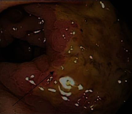
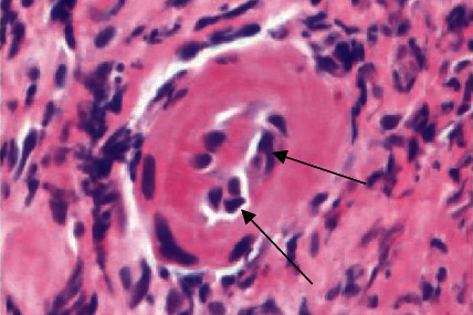
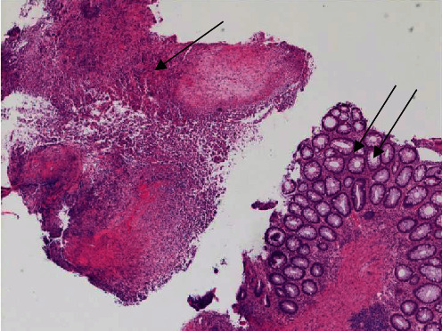
We describe two cases of rectal herpes simplex virus (HSV) infection in men that clinically mimicked rectal adenocarcinoma. Herpes infection in this location more commonly presents as an anal mass with viral inclusions in squamous epithelial cells. We report these cases to increase awareness of the unusual presentation as a proximal rectal mass with viral inclusions in endothelial cell nuclei. One patient was HIV-positive, and the other one had a history of having sex with men (MSM). Both patients had a thickened rectal wall with prominent lymphadenopathy on computed tomography (CT) scan, suspecting for malignancy. Biopsy showed abundant granulation tissue, necrosis, and inflammatory infiltrate composed predominantly of lymphocytes with admixed numerous plasma cells, eosinophils, and neutrophils. Rare granulation tissue vessels were lined by endothelial cells with nuclear molding and chromatin margination, and nuclei that were positive for HSV immunohistochemistry (IHC). One patient had confirmatory viral culture from biopsy of the ulcerated rectal mass. Both patients had symptom resolution following treatment for HSV. HSV should be considered in the differential diagnosis of rectal inflammatory masses, particularly in immunocompromised, HIV-positive, and MSM patients.


 分享
分享
 求助内容:
求助内容: 应助结果提醒方式:
 扫码关注我们
