{"title":"The Effect of Changes in the Angular Position of Implants on Metal Artifact Reduction in Cone-Beam Computed Tomography Images: A Scoping Review.","authors":"Maedeh Asnaashari, Maryam Sadeghipour, Zeinab Bahrani, Solmaz Valizadeh, Mahkameh Moshfeghi","doi":"10.1155/2023/5539719","DOIUrl":null,"url":null,"abstract":"<p><strong>Objective: </strong>Dental implant artifacts can compromise the quality of cone-beam computed tomography (CBCT) scans and challenge radiographic detection in surrounding regions. This literature review was conducted to examine the impact of implant angle modification on reducing metal artifacts in CBCT scans.</p><p><strong>Materials and methods: </strong>A scoping review of literature was carried out in PubMed, Embase, Scopus, and Cochrane databases.</p><p><strong>Results: </strong>Different spatial planes, including alpha, beta, gamma, and phi, along with 0°, 5.2°, 9.8°, 14.5°, 15°, 30°, 45°, 60°, 75°, and 90° angles were studied. Changes in the angular position of implants may reduce metal artifacts and improve the quality of CBCT scans.</p><p><strong>Conclusions: </strong>Rotating implants within the alpha plane and angling them at 90° in the alpha plane enables reducing dental implant artifacts.</p>","PeriodicalId":51864,"journal":{"name":"Radiology Research and Practice","volume":"2023 ","pages":"5539719"},"PeriodicalIF":1.5000,"publicationDate":"2023-07-31","publicationTypes":"Journal Article","fieldsOfStudy":null,"isOpenAccess":false,"openAccessPdf":"https://www.ncbi.nlm.nih.gov/pmc/articles/PMC10406552/pdf/","citationCount":"0","resultStr":null,"platform":"Semanticscholar","paperid":null,"PeriodicalName":"Radiology Research and Practice","FirstCategoryId":"1085","ListUrlMain":"https://doi.org/10.1155/2023/5539719","RegionNum":0,"RegionCategory":null,"ArticlePicture":[],"TitleCN":null,"AbstractTextCN":null,"PMCID":null,"EPubDate":"2023/1/1 0:00:00","PubModel":"eCollection","JCR":"Q2","JCRName":"RADIOLOGY, NUCLEAR MEDICINE & MEDICAL IMAGING","Score":null,"Total":0}
引用次数: 0
Abstract
Objective: Dental implant artifacts can compromise the quality of cone-beam computed tomography (CBCT) scans and challenge radiographic detection in surrounding regions. This literature review was conducted to examine the impact of implant angle modification on reducing metal artifacts in CBCT scans.
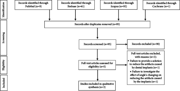
Materials and methods: A scoping review of literature was carried out in PubMed, Embase, Scopus, and Cochrane databases.
Results: Different spatial planes, including alpha, beta, gamma, and phi, along with 0°, 5.2°, 9.8°, 14.5°, 15°, 30°, 45°, 60°, 75°, and 90° angles were studied. Changes in the angular position of implants may reduce metal artifacts and improve the quality of CBCT scans.
Conclusions: Rotating implants within the alpha plane and angling them at 90° in the alpha plane enables reducing dental implant artifacts.
期刊介绍:
Radiology Research and Practice is a peer-reviewed, Open Access journal that publishes articles on all areas of medical imaging. The journal promotes evidence-based radiology practice though the publication of original research, reviews, and clinical studies for a multidisciplinary audience. Radiology Research and Practice is archived in Portico, which provides permanent archiving for electronic scholarly journals, as well as via the LOCKSS initiative. It operates a fully open access publishing model which allows open global access to its published content. This model is supported through Article Processing Charges. For more information on Article Processing charges in gen


 分享
分享
 求助内容:
求助内容: 应助结果提醒方式:
 扫码关注我们
