Osman Adi, Chan Pei Fong, Madeleine Kho Huei Tze, Azma Haryaty Ahmad, Nova Panebianco, Asri Ranga
{"title":"Transesophageal echocardiography (TEE)-guided transvenous pacing (TVP) in emergency department.","authors":"Osman Adi, Chan Pei Fong, Madeleine Kho Huei Tze, Azma Haryaty Ahmad, Nova Panebianco, Asri Ranga","doi":"10.1186/s13089-023-00332-7","DOIUrl":null,"url":null,"abstract":"<p><strong>Background: </strong>Placement of a temporary pacemaker is a vital skill in the emergency setting in patients that present with life-threatening bradycardia. Transvenous pacing is the definitive method of stabilizing the arrhythmia compared to transcutaneous pacing, as it provides more comfort and better control of heart rate, until the insertion of a permanent pacemaker.</p><p><strong>Case report: </strong>In this case report, we describe the steps using TEE to guide the insertion of transvenous pacer at the emergency department. Traditionally, the process of floating a transvenous pacer wire is performed \"blindly\" using landmarks and a monitoring ECG finding for capture, or under transthoracic echocardiography (TTE) ultrasound guidance. The blind procedure is associated with higher rate of failure and complications. While guidance using TTE is associated with higher success rates and fewer complications, inadequate imaging of the right side of the heart may limit the utility of this imaging modality. The use of transesophageal echocardiography (TEE) by emergency medicine and critical care physicians has gained traction in recent years due to its clear images and lack of interference with procedures being performed on the chest. In this article, we describe a protocol using TEE to guide the insertion of transvenous pacer through a case illustration.</p>","PeriodicalId":75201,"journal":{"name":"","volume":"15 1","pages":"35"},"PeriodicalIF":0.0,"publicationDate":"2023-08-21","publicationTypes":"Journal Article","fieldsOfStudy":null,"isOpenAccess":false,"openAccessPdf":"https://www.ncbi.nlm.nih.gov/pmc/articles/PMC10441836/pdf/","citationCount":"0","resultStr":null,"platform":"Semanticscholar","paperid":null,"PeriodicalName":"","FirstCategoryId":"1085","ListUrlMain":"https://doi.org/10.1186/s13089-023-00332-7","RegionNum":0,"RegionCategory":null,"ArticlePicture":[],"TitleCN":null,"AbstractTextCN":null,"PMCID":null,"EPubDate":"","PubModel":"","JCR":"","JCRName":"","Score":null,"Total":0}
引用次数: 0
Abstract
Background: Placement of a temporary pacemaker is a vital skill in the emergency setting in patients that present with life-threatening bradycardia. Transvenous pacing is the definitive method of stabilizing the arrhythmia compared to transcutaneous pacing, as it provides more comfort and better control of heart rate, until the insertion of a permanent pacemaker.
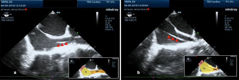
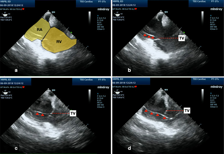
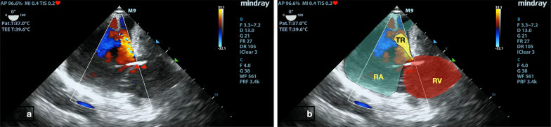
Case report: In this case report, we describe the steps using TEE to guide the insertion of transvenous pacer at the emergency department. Traditionally, the process of floating a transvenous pacer wire is performed "blindly" using landmarks and a monitoring ECG finding for capture, or under transthoracic echocardiography (TTE) ultrasound guidance. The blind procedure is associated with higher rate of failure and complications. While guidance using TTE is associated with higher success rates and fewer complications, inadequate imaging of the right side of the heart may limit the utility of this imaging modality. The use of transesophageal echocardiography (TEE) by emergency medicine and critical care physicians has gained traction in recent years due to its clear images and lack of interference with procedures being performed on the chest. In this article, we describe a protocol using TEE to guide the insertion of transvenous pacer through a case illustration.


 分享
分享
 求助内容:
求助内容: 应助结果提醒方式:
 扫码关注我们
