Jacob B Pears, Carley Tillett, Rui Tahara, Hans C E Larsson, Kate Trinajstic, Catherine A Boisvert
{"title":"The Development of the <i>Chimaeroid</i> Pelvic Skeleton and the Evolution of Chondrichthyan Pelvic Fins.","authors":"Jacob B Pears, Carley Tillett, Rui Tahara, Hans C E Larsson, Kate Trinajstic, Catherine A Boisvert","doi":"10.3390/jdb10040053","DOIUrl":null,"url":null,"abstract":"<p><p>Pelvic girdles, fins and claspers are evolutionary novelties first recorded in jawed vertebrates. Over the course of the evolution of chondrichthyans (cartilaginous fish) two trends in the morphology of the pelvic skeleton have been suggested to have occurred. These evolutionary shifts involved both an enlargement of the metapterygium (basipterygium) and a transition of fin radial articulation from the pelvic girdle to the metapterygium. To determine how these changes in morphology have occurred it is essential to understand the development of extant taxa as this can indicate potential developmental mechanisms that may have been responsible for these changes. The study of the morphology of the appendicular skeleton across development in chondrichthyans is almost entirely restricted to the historical literature with little contemporary research. Here, we have examined the morphology and development of the pelvic skeleton of a holocephalan chondrichthyan, the elephant shark (<i>Callorhinchus milii</i>), through a combination of dissections, histology, and nanoCT imaging and redescribed the pelvic skeleton of <i>Cladoselache kepleri</i> (NHMUK PV P 9269), a stem holocephalan. To put our findings in their evolutionary context we compare them with the fossil record of chondrichthyans and the literature on pelvic development in elasmobranchs from the late 19th century. Our findings demonstrate that the pelvic skeleton of <i>C. milii</i> initially forms as a single mesenchymal condensation, consisting of the pelvic girdle and a series of fin rays, which fuse to form the basipterygium. The girdle and fin skeleton subsequently segment into distinct components whilst chondrifying. This confirms descriptions of the early pelvic development in <i>Scyliorhinid</i> sharks from the historical literature and suggests that chimaeras and elasmobranchs share common developmental patterns in their pelvic anatomy. Alterations in the location and degree of radial fusion during early development may be the mechanism responsible for changes in pelvic fin morphology over the course of the evolution of both elasmobranchs and holocephalans, which appears to be an example of parallel evolution.</p>","PeriodicalId":15563,"journal":{"name":"Journal of Developmental Biology","volume":"10 4","pages":""},"PeriodicalIF":2.5000,"publicationDate":"2022-12-12","publicationTypes":"Journal Article","fieldsOfStudy":null,"isOpenAccess":false,"openAccessPdf":"https://www.ncbi.nlm.nih.gov/pmc/articles/PMC9782884/pdf/","citationCount":"0","resultStr":null,"platform":"Semanticscholar","paperid":null,"PeriodicalName":"Journal of Developmental Biology","FirstCategoryId":"1085","ListUrlMain":"https://doi.org/10.3390/jdb10040053","RegionNum":0,"RegionCategory":null,"ArticlePicture":[],"TitleCN":null,"AbstractTextCN":null,"PMCID":null,"EPubDate":"","PubModel":"","JCR":"Q3","JCRName":"DEVELOPMENTAL BIOLOGY","Score":null,"Total":0}
引用次数: 0
Abstract
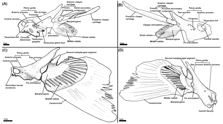
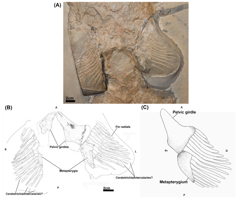
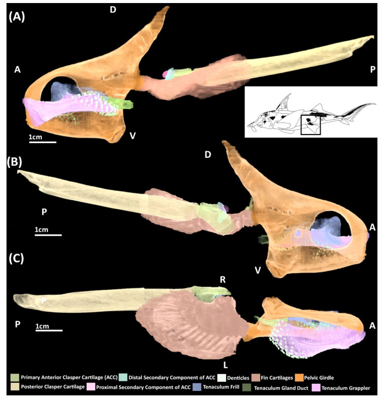
Pelvic girdles, fins and claspers are evolutionary novelties first recorded in jawed vertebrates. Over the course of the evolution of chondrichthyans (cartilaginous fish) two trends in the morphology of the pelvic skeleton have been suggested to have occurred. These evolutionary shifts involved both an enlargement of the metapterygium (basipterygium) and a transition of fin radial articulation from the pelvic girdle to the metapterygium. To determine how these changes in morphology have occurred it is essential to understand the development of extant taxa as this can indicate potential developmental mechanisms that may have been responsible for these changes. The study of the morphology of the appendicular skeleton across development in chondrichthyans is almost entirely restricted to the historical literature with little contemporary research. Here, we have examined the morphology and development of the pelvic skeleton of a holocephalan chondrichthyan, the elephant shark (Callorhinchus milii), through a combination of dissections, histology, and nanoCT imaging and redescribed the pelvic skeleton of Cladoselache kepleri (NHMUK PV P 9269), a stem holocephalan. To put our findings in their evolutionary context we compare them with the fossil record of chondrichthyans and the literature on pelvic development in elasmobranchs from the late 19th century. Our findings demonstrate that the pelvic skeleton of C. milii initially forms as a single mesenchymal condensation, consisting of the pelvic girdle and a series of fin rays, which fuse to form the basipterygium. The girdle and fin skeleton subsequently segment into distinct components whilst chondrifying. This confirms descriptions of the early pelvic development in Scyliorhinid sharks from the historical literature and suggests that chimaeras and elasmobranchs share common developmental patterns in their pelvic anatomy. Alterations in the location and degree of radial fusion during early development may be the mechanism responsible for changes in pelvic fin morphology over the course of the evolution of both elasmobranchs and holocephalans, which appears to be an example of parallel evolution.
期刊介绍:
The Journal of Developmental Biology (ISSN 2221-3759) is an international, peer-reviewed, quick-refereeing, open access journal, which publishes reviews, research papers and communications on the development of multicellular organisms at the molecule, cell, tissue, organ and whole organism levels. Our aim is to encourage researchers to effortlessly publish their new findings or concepts rapidly in an open access medium, overseen by their peers. There is no restriction on the length of the papers; the full experimental details must be provided so that the results can be reproduced. Electronic files regarding the full details of the experimental procedure, if unable to be published in a normal way, can be deposited as supplementary material. Journal of Developmental Biology focuses on: -Development mechanisms and genetics -Cell differentiation -Embryonal development -Tissue/organism growth -Metamorphosis and regeneration of the organisms. It involves many biological fields, such as Molecular biology, Genetics, Physiology, Cell biology, Anatomy, Embryology, Cancer research, Neurobiology, Immunology, Ecology, Evolutionary biology.


 分享
分享
 求助内容:
求助内容: 应助结果提醒方式:
 扫码关注我们
