Nan Liu, Yi Guan, Yan Yu, Gai Li, Ling Xue, Weikang Li, Xiaoyu Qu, Ning Li, Sanqiao Yao
{"title":"Pulmonary effects of exposure to indium and its compounds: cross-sectional survey of exposed workers and experimental findings in rodents.","authors":"Nan Liu, Yi Guan, Yan Yu, Gai Li, Ling Xue, Weikang Li, Xiaoyu Qu, Ning Li, Sanqiao Yao","doi":"10.1186/s12989-022-00510-w","DOIUrl":null,"url":null,"abstract":"<p><strong>Background: </strong>Many studies have shown that occupational exposure to indium and its compounds could induce lung disease. Although animal toxicological studies and human epidemiological studies suggest indium exposure may cause lung injury, inflammation, pulmonary fibrosis, emphysema, pulmonary alveolar proteinosis, and even lung cancer, related data collected from humans is currently limited and confined to single workplaces, and the early effects of exposure on the lungs are not well understood.</p><p><strong>Objectives: </strong>This study combined population studies and animal experiments to examine the links of indium with pulmonary injury, as well as its mechanism of action. A cross-sectional epidemiological study of indium-exposed workers from China was conducted to evaluate associations between occupational indium exposure and serum biomarkers of early effect. This study also compares and analyzes the causal perspectives of changes in human serum biomarkers induced by indium compound exposure and indium exposure-related rat lung pathobiology, and discusses possible avenues for their recognition and prevention.</p><p><strong>Methods: </strong>This is a study of 57 exposed (at least 6 h per day for one year) workers from an indium ingot production plant, and 63 controls. Indium concentration in serum, urine, and airborne as exposure indices were measured by inductively coupled plasma-mass spectrometry. Sixteen serum biomarkers of pulmonary injury, inflammation, and oxidative stress were measured using ELISA. The associations between serum indium and 16 serum biomarkers were analyzed to explore the mechanism of action of indium on pulmonary injury in indium-exposed workers. Animal experiments were conducted to measure inflammatory factors levels in bronchoalveolar lavage fluid (BALF) and lung tissue protein expressions in rats. Four different forms of indium compound-exposed rat models were established (intratracheal instillation twice per week, 8 week exposure, 8 week recovery). Model I: 0, 1.2, 3, and 6 mg/kg bw indium tin oxide group; Model II: 0, 1.2, 3, and 6 mg/kg bw indium oxide (In<sub>2</sub>O<sub>3</sub>) group; Model III: 0, 0.523, 1.046, and 2.614 mg/kg bw indium sulfate (In<sub>2</sub>(SO<sub>4</sub>)<sub>3</sub>) group; Model IV: 0, 0.065, 0.65, and 1.3 mg/kg bw indium trichloride (InCl<sub>3</sub>) group. Lung pathological changes were assessed by hematoxylin & eosin, periodic acid Schiff, and Masson's staining, transmission electron microscopy, and the protein changes were determined by immunohistochemistry.</p><p><strong>Results: </strong>In the production workshop, the airborne indium concentration was 78.4 μg/m<sup>3</sup>. The levels of serum indium and urine indium in indium-exposed workers were 39.3 μg/L and 11.0 ng/g creatinine. Increased lung damage markers, oxidative stress markers, and inflammation markers were found in indium-exposed workers. Serum indium levels were statistically and positively associated with the serum levels of SP-A, IL-1β, IL-6 in indium-exposed workers. Among them, SP-A showed a duration-response pattern. The results of animal experiments showed that, with an increase in dosage, indium exposure significantly increased the levels of serum indium and lung indium, as well as the BALF levels of IL‑1β, IL‑6, IL‑10, and TNF‑α and up-regulated the protein expression of SP-A, SP-D, KL-6, GM-CSF, NF-κB p65, and HO-1 in all rat models groups. TEM revealed that In<sub>2</sub>(SO<sub>4</sub>)<sub>3</sub> and InCl<sub>3</sub> are soluble and that no particles were found in lung tissue, in contrast to the non-soluble compounds (ITO and In<sub>2</sub>O<sub>3</sub>). No PAS-staining positive substance was found in the lung tissue of In<sub>2</sub>(SO<sub>4</sub>)<sub>3</sub> and InCl<sub>3</sub> exposure groups, whereas ITO and In<sub>2</sub>O<sub>3</sub> rat models supported findings of pulmonary alveolar proteinosis and interstitial fibrosis seen in human indium lung disease. ITO and InCl<sub>3</sub> can accelerate interstitial fibrosis. Findings from our in vivo studies demonstrated that intra-alveolar accumulation of surfactant (immunohistochemistry) and characteristic cholesterol clefts granulomas of indium lung disease (PAS staining) were triggered by a specific form of indium (ITO and In<sub>2</sub>O<sub>3</sub>).</p><p><strong>Conclusions: </strong>In indium-exposed workers, biomarker findings indicated lung damage, oxidative stress and an inflammatory response. In rat models of the four forms of indium encountered in a workplace, the biomarkers response to all compounds overall corresponded to that in humans. In addition, pulmonary alveolar proteinosis was found following exposure to indium tin oxide and indium oxide in the rat models, and interstitial fibrosis was found following exposure to indium tin oxide and indium trichloride, supporting previous report of human disease. Serum SP-A levels were positively associated with indium exposure and may be considered a potential biomarker of exposure and effect in exposed workers.</p>","PeriodicalId":19847,"journal":{"name":"Particle and Fibre Toxicology","volume":"19 1","pages":"69"},"PeriodicalIF":8.2000,"publicationDate":"2022-12-20","publicationTypes":"Journal Article","fieldsOfStudy":null,"isOpenAccess":false,"openAccessPdf":"https://www.ncbi.nlm.nih.gov/pmc/articles/PMC9764635/pdf/","citationCount":"0","resultStr":null,"platform":"Semanticscholar","paperid":null,"PeriodicalName":"Particle and Fibre Toxicology","FirstCategoryId":"3","ListUrlMain":"https://doi.org/10.1186/s12989-022-00510-w","RegionNum":1,"RegionCategory":"医学","ArticlePicture":[],"TitleCN":null,"AbstractTextCN":null,"PMCID":null,"EPubDate":"","PubModel":"","JCR":"Q1","JCRName":"TOXICOLOGY","Score":null,"Total":0}
引用次数: 0
Abstract
Background: Many studies have shown that occupational exposure to indium and its compounds could induce lung disease. Although animal toxicological studies and human epidemiological studies suggest indium exposure may cause lung injury, inflammation, pulmonary fibrosis, emphysema, pulmonary alveolar proteinosis, and even lung cancer, related data collected from humans is currently limited and confined to single workplaces, and the early effects of exposure on the lungs are not well understood.
Objectives: This study combined population studies and animal experiments to examine the links of indium with pulmonary injury, as well as its mechanism of action. A cross-sectional epidemiological study of indium-exposed workers from China was conducted to evaluate associations between occupational indium exposure and serum biomarkers of early effect. This study also compares and analyzes the causal perspectives of changes in human serum biomarkers induced by indium compound exposure and indium exposure-related rat lung pathobiology, and discusses possible avenues for their recognition and prevention.
Methods: This is a study of 57 exposed (at least 6 h per day for one year) workers from an indium ingot production plant, and 63 controls. Indium concentration in serum, urine, and airborne as exposure indices were measured by inductively coupled plasma-mass spectrometry. Sixteen serum biomarkers of pulmonary injury, inflammation, and oxidative stress were measured using ELISA. The associations between serum indium and 16 serum biomarkers were analyzed to explore the mechanism of action of indium on pulmonary injury in indium-exposed workers. Animal experiments were conducted to measure inflammatory factors levels in bronchoalveolar lavage fluid (BALF) and lung tissue protein expressions in rats. Four different forms of indium compound-exposed rat models were established (intratracheal instillation twice per week, 8 week exposure, 8 week recovery). Model I: 0, 1.2, 3, and 6 mg/kg bw indium tin oxide group; Model II: 0, 1.2, 3, and 6 mg/kg bw indium oxide (In2O3) group; Model III: 0, 0.523, 1.046, and 2.614 mg/kg bw indium sulfate (In2(SO4)3) group; Model IV: 0, 0.065, 0.65, and 1.3 mg/kg bw indium trichloride (InCl3) group. Lung pathological changes were assessed by hematoxylin & eosin, periodic acid Schiff, and Masson's staining, transmission electron microscopy, and the protein changes were determined by immunohistochemistry.
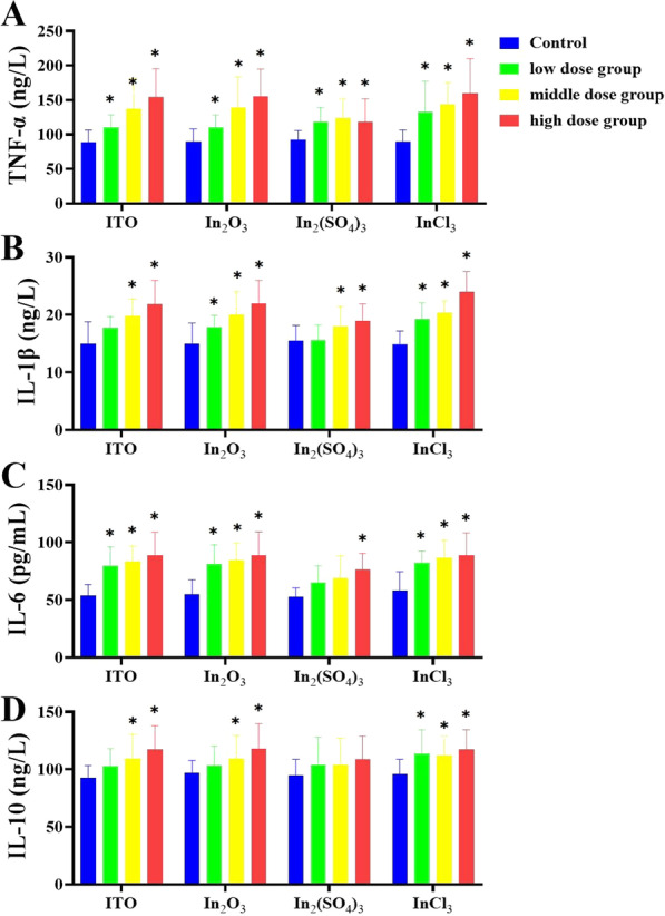
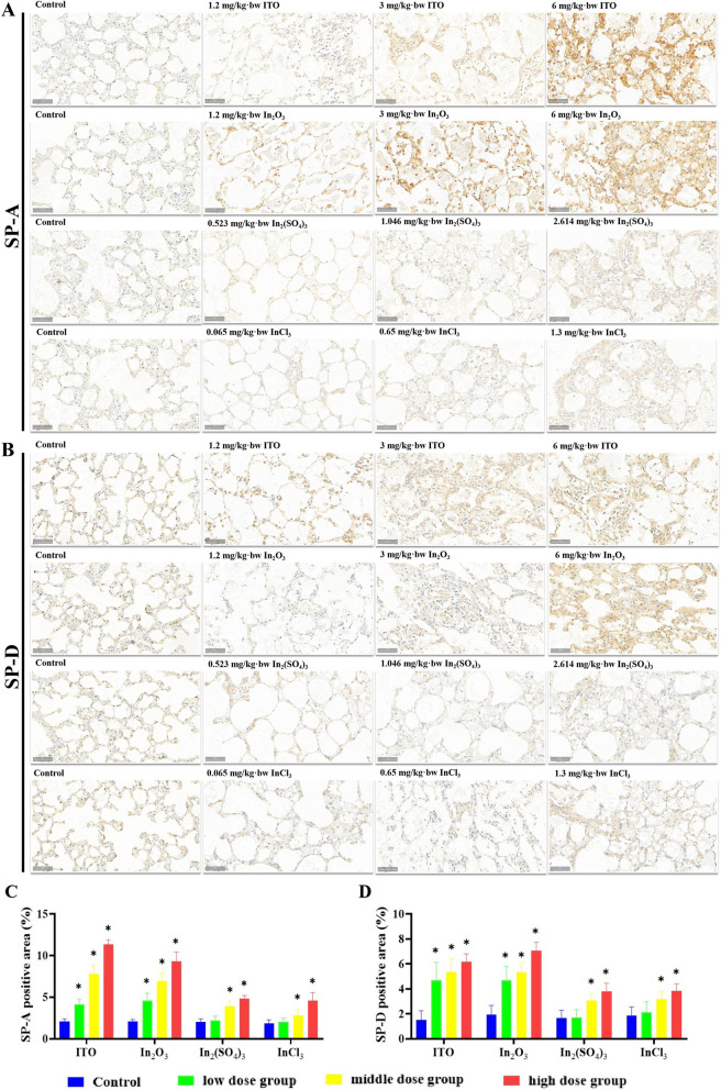
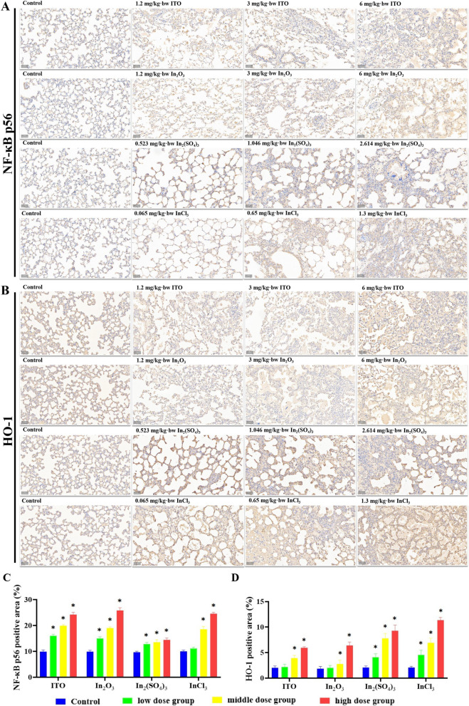
Results: In the production workshop, the airborne indium concentration was 78.4 μg/m3. The levels of serum indium and urine indium in indium-exposed workers were 39.3 μg/L and 11.0 ng/g creatinine. Increased lung damage markers, oxidative stress markers, and inflammation markers were found in indium-exposed workers. Serum indium levels were statistically and positively associated with the serum levels of SP-A, IL-1β, IL-6 in indium-exposed workers. Among them, SP-A showed a duration-response pattern. The results of animal experiments showed that, with an increase in dosage, indium exposure significantly increased the levels of serum indium and lung indium, as well as the BALF levels of IL‑1β, IL‑6, IL‑10, and TNF‑α and up-regulated the protein expression of SP-A, SP-D, KL-6, GM-CSF, NF-κB p65, and HO-1 in all rat models groups. TEM revealed that In2(SO4)3 and InCl3 are soluble and that no particles were found in lung tissue, in contrast to the non-soluble compounds (ITO and In2O3). No PAS-staining positive substance was found in the lung tissue of In2(SO4)3 and InCl3 exposure groups, whereas ITO and In2O3 rat models supported findings of pulmonary alveolar proteinosis and interstitial fibrosis seen in human indium lung disease. ITO and InCl3 can accelerate interstitial fibrosis. Findings from our in vivo studies demonstrated that intra-alveolar accumulation of surfactant (immunohistochemistry) and characteristic cholesterol clefts granulomas of indium lung disease (PAS staining) were triggered by a specific form of indium (ITO and In2O3).
Conclusions: In indium-exposed workers, biomarker findings indicated lung damage, oxidative stress and an inflammatory response. In rat models of the four forms of indium encountered in a workplace, the biomarkers response to all compounds overall corresponded to that in humans. In addition, pulmonary alveolar proteinosis was found following exposure to indium tin oxide and indium oxide in the rat models, and interstitial fibrosis was found following exposure to indium tin oxide and indium trichloride, supporting previous report of human disease. Serum SP-A levels were positively associated with indium exposure and may be considered a potential biomarker of exposure and effect in exposed workers.
期刊介绍:
Particle and Fibre Toxicology is an online journal that is open access and peer-reviewed. It covers a range of disciplines such as material science, biomaterials, and nanomedicine, focusing on the toxicological effects of particles and fibres. The journal serves as a platform for scientific debate and communication among toxicologists and scientists from different fields who work with particle and fibre materials. The main objective of the journal is to deepen our understanding of the physico-chemical properties of particles, their potential for human exposure, and the resulting biological effects. It also addresses regulatory issues related to particle exposure in workplaces and the general environment. Moreover, the journal recognizes that there are various situations where particles can pose a toxicological threat, such as the use of old materials in new applications or the introduction of new materials altogether. By encompassing all these disciplines, Particle and Fibre Toxicology provides a comprehensive source for research in this field.


 分享
分享
 求助内容:
求助内容: 应助结果提醒方式:
 扫码关注我们
