{"title":"Co-Culture of Mouse Blastocysts on A Human Recellularized Endometrial Scaffold: An <i>In Vitro</i> Model for Future Implantation Studies.","authors":"Elham Sadeghi, Mojtaba Rezazadeh Valojerdi, Mojdeh Salehnia","doi":"10.22074/cellj.2023.1989926.1236","DOIUrl":null,"url":null,"abstract":"<p><strong>Objective: </strong>This study evaluates the interaction of mouse blastocysts as a surrogate embryo on a recellularized endometrial scaffold by seeding human endometrial mesenchymal cells (hEMCs).</p><p><strong>Materials and methods: </strong>In this experimental study, prepared decellularized human endometrial tissues were characterized by morphological staining, DNA content analysis, and scanning electron microscopic (SEM) analysis. The scaffolds were subsequently recellularized by hEMCs. After seven days of cultivation, the mouse blastocysts were co-cultured on the recellularized scaffolds for 48 hours. Embryo attachment and implantation within these scaffolds were evaluated at the morphological, ultrastructural, molecular, and hormonal levels.</p><p><strong>Results: </strong>There was no morphological evidence of cells and nuclei in the decellularized scaffold. DNA content significantly decreased by 89.92% compared to the control group (P<0.05). Both decellularized and native tissues had similar patterns of collagen bundles and elastin fibers, and glycosaminoglycan (GAGs) distribution in the stroma. After recellularization, the hEMCs attached to the scaffold surface and penetrated different parts of these scaffolds. In the co-cultured group, the embryo attached to the surface of the scaffold after 24 hours and penetrated the recellularized endometrial tissue after 48 hours. We observed multi-layered organoid-like structures formed by hEMC proliferation. The relative expressions of epithelial-related genes, <i>ZO-1</i> and <i>COL4A1</i>, and <i>SSP1, MMP2,</i> and <i>PRL</i>, as decidualizationrelated genes, were significantly higher in the recellularized group on day 9 in the presence of the embryo compared to the other groups (P<0.05). Beta human chorionic gonadotropin (β-hCG) and prolactin were statistically increased in the recellularized group on day 9 group (P<0.05).</p><p><strong>Conclusion: </strong>hEMCs and mouse embryo co-cultured on a decellularized endometrial scaffold provides an alternative model to study embryo implantation and the earlier stage of embryo development.</p>","PeriodicalId":49224,"journal":{"name":"Cell Journal","volume":"25 8","pages":"579-590"},"PeriodicalIF":1.7000,"publicationDate":"2023-08-01","publicationTypes":"Journal Article","fieldsOfStudy":null,"isOpenAccess":false,"openAccessPdf":"https://ftp.ncbi.nlm.nih.gov/pub/pmc/oa_pdf/b2/0b/Cell-J-25-579.PMC10542203.pdf","citationCount":"0","resultStr":null,"platform":"Semanticscholar","paperid":null,"PeriodicalName":"Cell Journal","FirstCategoryId":"99","ListUrlMain":"https://doi.org/10.22074/cellj.2023.1989926.1236","RegionNum":4,"RegionCategory":"生物学","ArticlePicture":[],"TitleCN":null,"AbstractTextCN":null,"PMCID":null,"EPubDate":"","PubModel":"","JCR":"Q4","JCRName":"CELL BIOLOGY","Score":null,"Total":0}
引用次数: 0
Abstract
Objective: This study evaluates the interaction of mouse blastocysts as a surrogate embryo on a recellularized endometrial scaffold by seeding human endometrial mesenchymal cells (hEMCs).
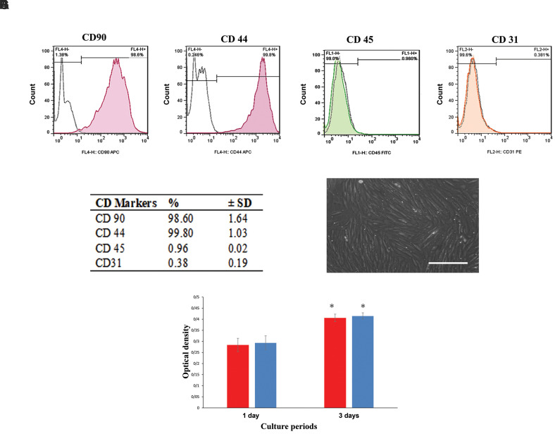
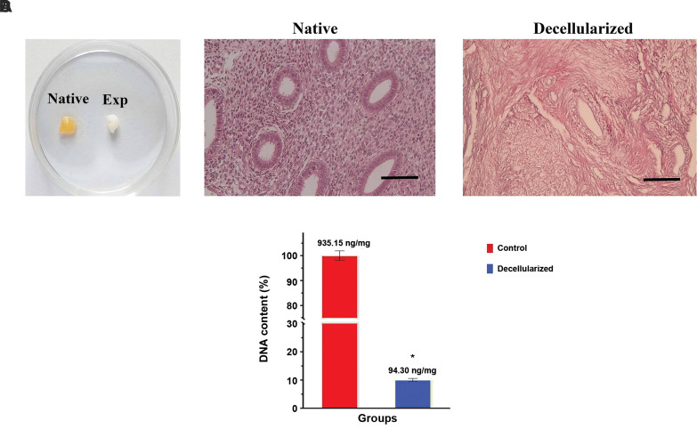
Materials and methods: In this experimental study, prepared decellularized human endometrial tissues were characterized by morphological staining, DNA content analysis, and scanning electron microscopic (SEM) analysis. The scaffolds were subsequently recellularized by hEMCs. After seven days of cultivation, the mouse blastocysts were co-cultured on the recellularized scaffolds for 48 hours. Embryo attachment and implantation within these scaffolds were evaluated at the morphological, ultrastructural, molecular, and hormonal levels.
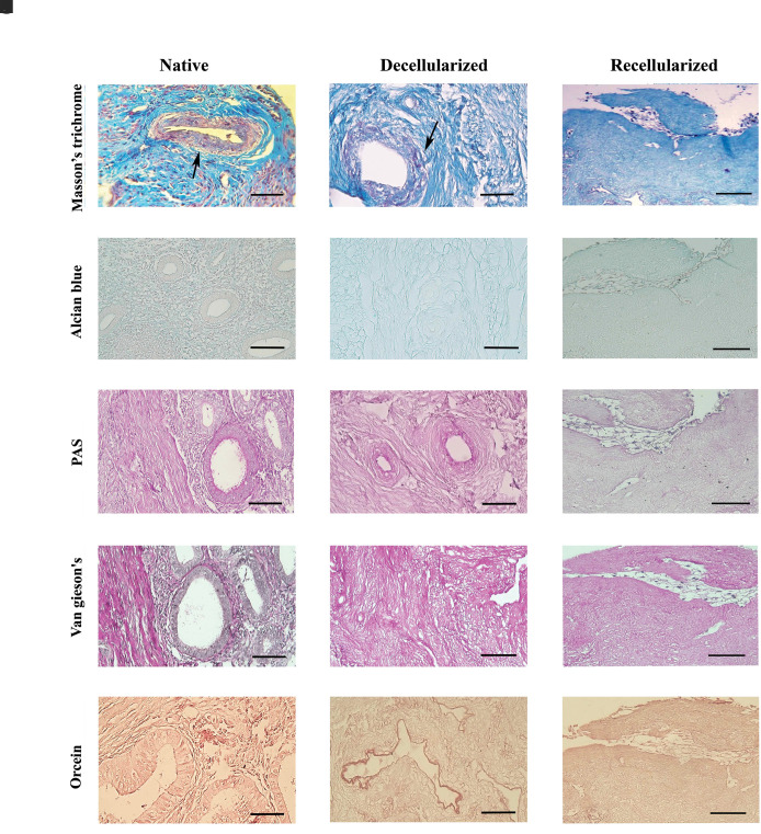
Results: There was no morphological evidence of cells and nuclei in the decellularized scaffold. DNA content significantly decreased by 89.92% compared to the control group (P<0.05). Both decellularized and native tissues had similar patterns of collagen bundles and elastin fibers, and glycosaminoglycan (GAGs) distribution in the stroma. After recellularization, the hEMCs attached to the scaffold surface and penetrated different parts of these scaffolds. In the co-cultured group, the embryo attached to the surface of the scaffold after 24 hours and penetrated the recellularized endometrial tissue after 48 hours. We observed multi-layered organoid-like structures formed by hEMC proliferation. The relative expressions of epithelial-related genes, ZO-1 and COL4A1, and SSP1, MMP2, and PRL, as decidualizationrelated genes, were significantly higher in the recellularized group on day 9 in the presence of the embryo compared to the other groups (P<0.05). Beta human chorionic gonadotropin (β-hCG) and prolactin were statistically increased in the recellularized group on day 9 group (P<0.05).
Conclusion: hEMCs and mouse embryo co-cultured on a decellularized endometrial scaffold provides an alternative model to study embryo implantation and the earlier stage of embryo development.
期刊介绍:
The “Cell Journal (Yakhteh)“, formerly published as “Yakhteh Medical Journal”, is a quarterly English publication of Royan Institute. This journal focuses on topics relevant to cellular and molecular scientific areas, besides other related fields. The Cell J has been certified by Ministry of Culture and Islamic Guidance in 1999 and was accredited as a scientific and research journal by HBI (Health and Biomedical Information) Journal Accreditation Commission in 2000 which is an open access journal.


 分享
分享
 求助内容:
求助内容: 应助结果提醒方式:
 扫码关注我们
