Mostafa EmadEldeen Hussien Mohamed Afify, Randa Hesham Ali Abdelgawad, Momen Mahmoud Hamdi, Amany Abd El-Fattah El-Shazly, Mohamed Adel Abdelshafik
{"title":"Multifocal visual evoked potential for evaluation of open-angle glaucoma.","authors":"Mostafa EmadEldeen Hussien Mohamed Afify, Randa Hesham Ali Abdelgawad, Momen Mahmoud Hamdi, Amany Abd El-Fattah El-Shazly, Mohamed Adel Abdelshafik","doi":"10.51329/mehdiophthal1429","DOIUrl":null,"url":null,"abstract":"<p><strong>Background: </strong>To correlate multifocal visual evoked potential (mfVEP) findings with static automated perimetry (SAP) and spectral-domain optical coherence tomography (SD-OCT) in eyes with primary open- angle glaucoma (POAG).</p><p><strong>Methods: </strong>This cross-sectional study included a consecutive sample of 40 eyes of 40 patients with POAG. The participants underwent a complete ophthalmologic assessment, axial length (AL) measurement, and assessments with SAP, SD-OCT, and mfVEP.</p><p><strong>Results: </strong>POAG cases were aged 49.70 (14.16) years (mean [SD]) and most were females (n = 24, 60%). For eyes of patients with POAG, the mfVEP upper-ring signal-to-noise ratio (SNR) showed a significant negative correlation with best-corrected logMAR visual acuity (r = - 0.33; <i>P</i> = 0.038), and a significant positive correlation with the superior hemifield of the visual field (VF) and the inferior-quadrant retinal nerve fiber layer (RNFL) thickness (r = + 0.34; <i>P</i> = 0.030; r = + 0.51; <i>P</i> < 0.001, respectively). Similarly, the mfVEP lower-ring SNR showed a significant negative correlation with best-corrected logMAR visual acuity (r = - 0.36; <i>P</i> = 0.024) and a significant positive correlation with the inferior hemifield of the VF and superior quadrant RNFL thickness (r = + 0.55; <i>P</i> < 0.001 and r = + 0.70; <i>P</i> < 0.001, respectively).</p><p><strong>Conclusions: </strong>mfVEP is a promising tool for objective assessment of the VF in patients with POAG, as it is positively correlated with the VF and OCT RNFL thickness. Future longitudinal studies with a larger sample size and a specific glaucoma subtype, along with multiple follow-up evaluations, are warranted to confirm our preliminary results.</p>","PeriodicalId":36524,"journal":{"name":"Medical Hypothesis, Discovery, and Innovation in Ophthalmology","volume":"10 3","pages":"114-120"},"PeriodicalIF":0.0000,"publicationDate":"2021-01-01","publicationTypes":"Journal Article","fieldsOfStudy":null,"isOpenAccess":false,"openAccessPdf":"https://ftp.ncbi.nlm.nih.gov/pub/pmc/oa_pdf/ab/9b/mehdiophth-10-114.PMC10460219.pdf","citationCount":"0","resultStr":null,"platform":"Semanticscholar","paperid":null,"PeriodicalName":"Medical Hypothesis, Discovery, and Innovation in Ophthalmology","FirstCategoryId":"1085","ListUrlMain":"https://doi.org/10.51329/mehdiophthal1429","RegionNum":0,"RegionCategory":null,"ArticlePicture":[],"TitleCN":null,"AbstractTextCN":null,"PMCID":null,"EPubDate":"","PubModel":"","JCR":"Q2","JCRName":"Medicine","Score":null,"Total":0}
引用次数: 0
Abstract
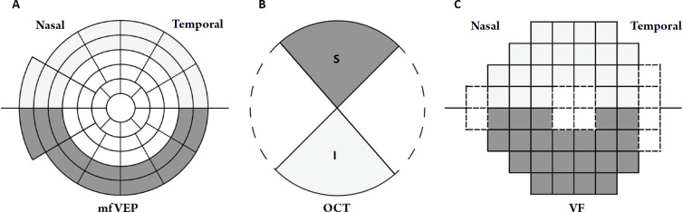
Background: To correlate multifocal visual evoked potential (mfVEP) findings with static automated perimetry (SAP) and spectral-domain optical coherence tomography (SD-OCT) in eyes with primary open- angle glaucoma (POAG).
Methods: This cross-sectional study included a consecutive sample of 40 eyes of 40 patients with POAG. The participants underwent a complete ophthalmologic assessment, axial length (AL) measurement, and assessments with SAP, SD-OCT, and mfVEP.
Results: POAG cases were aged 49.70 (14.16) years (mean [SD]) and most were females (n = 24, 60%). For eyes of patients with POAG, the mfVEP upper-ring signal-to-noise ratio (SNR) showed a significant negative correlation with best-corrected logMAR visual acuity (r = - 0.33; P = 0.038), and a significant positive correlation with the superior hemifield of the visual field (VF) and the inferior-quadrant retinal nerve fiber layer (RNFL) thickness (r = + 0.34; P = 0.030; r = + 0.51; P < 0.001, respectively). Similarly, the mfVEP lower-ring SNR showed a significant negative correlation with best-corrected logMAR visual acuity (r = - 0.36; P = 0.024) and a significant positive correlation with the inferior hemifield of the VF and superior quadrant RNFL thickness (r = + 0.55; P < 0.001 and r = + 0.70; P < 0.001, respectively).
Conclusions: mfVEP is a promising tool for objective assessment of the VF in patients with POAG, as it is positively correlated with the VF and OCT RNFL thickness. Future longitudinal studies with a larger sample size and a specific glaucoma subtype, along with multiple follow-up evaluations, are warranted to confirm our preliminary results.


 分享
分享
 求助内容:
求助内容: 应助结果提醒方式:
 扫码关注我们
