Mariam A Elshawarby, Ali Saad, Thanaa Helmy, Mouamen M Seleet, Tamer Elraggal
{"title":"Functional optical zone after wavefront-optimized versus wavefront-guided laser in situ keratomileusis.","authors":"Mariam A Elshawarby, Ali Saad, Thanaa Helmy, Mouamen M Seleet, Tamer Elraggal","doi":"10.51329/mehdiophthal1431","DOIUrl":null,"url":null,"abstract":"<p><strong>Background: </strong>Many studies have used functional optical zone (FOZ) as a measure to compare different refractive laser treatment modalities. However, to our knowledge, no study has compared wavefront- optimized (WFO) and wavefront-guided (WFG) laser in situ keratomileusis (LASIK) using FOZ. We compared the FOZ after WFO versus WFG LASIK in patients with myopia and myopic astigmatism.</p><p><strong>Methods: </strong>In this prospective comparative study, we included 100 myopic eyes of 50 patients with or without astigmatism. They were divided into two groups according to the platform used: WFO or WFG femtosecond LASIK. Using Holladay's equivalent keratometry reading (EKR) report of Pentacam HR, FOZ was defined as a zone centered on the pupil center with a standard deviation (SD) of 0.5 D, around the mean EKR. The differences in FOZ between the two platforms were analyzed at 3 months postoperatively. Visual acuity, refractive error, corneal asphericity (Q-value), and root mean square of higher-order aberrations (RMS for HOAs) were evaluated and compared.</p><p><strong>Results: </strong>The mean (SD) of patient age was 26.64 (5.67) years. The preoperative characteristics of the two groups were comparable (all <i>P</i> > 0.05). The intended optical zone (IOZ) was 6 mm in both groups. The mean laser ablation depth was significantly greater in the WFG group (18 µm per D) than in the WFO group (16 µm per D) (<i>P</i> = 0.035). At 3 months postoperatively, the mean (SD) of FOZ diameter was 4.32 (0.94) mm (71.99% [15.68%] of intended optical zone) in the WFO group and 4.16 (1.13) mm (69.33% [18.78%] of intended optical zone) in the WFG group, with no significant difference between the two groups (<i>P</i> = 0.622). The change in corneal asphericity was greater in the WFG group than in the WFO group (<i>P</i> = 0.034). Postoperative mean corrected and uncorrected distance visual acuity, manifest refraction, and RMS for HOAs showed no significant difference between the two groups (all <i>P</i> > 0.05).</p><p><strong>Conclusions: </strong>We found that WFG LASIK resulted in greater ablation depth and change in corneal asphericity than WFO LASIK at 3 months postoperatively. However, there was no significant difference in FOZ diameter, refractive error, and RMS for HOAs between the two groups. Further research is needed to confirm these findings.</p>","PeriodicalId":36524,"journal":{"name":"Medical Hypothesis, Discovery, and Innovation in Ophthalmology","volume":"10 3","pages":"129-137"},"PeriodicalIF":0.0000,"publicationDate":"2021-01-01","publicationTypes":"Journal Article","fieldsOfStudy":null,"isOpenAccess":false,"openAccessPdf":"https://ftp.ncbi.nlm.nih.gov/pub/pmc/oa_pdf/39/e3/mehdiophth-10-129.PMC10460224.pdf","citationCount":"0","resultStr":null,"platform":"Semanticscholar","paperid":null,"PeriodicalName":"Medical Hypothesis, Discovery, and Innovation in Ophthalmology","FirstCategoryId":"1085","ListUrlMain":"https://doi.org/10.51329/mehdiophthal1431","RegionNum":0,"RegionCategory":null,"ArticlePicture":[],"TitleCN":null,"AbstractTextCN":null,"PMCID":null,"EPubDate":"","PubModel":"","JCR":"Q2","JCRName":"Medicine","Score":null,"Total":0}
引用次数: 0
Abstract
Background: Many studies have used functional optical zone (FOZ) as a measure to compare different refractive laser treatment modalities. However, to our knowledge, no study has compared wavefront- optimized (WFO) and wavefront-guided (WFG) laser in situ keratomileusis (LASIK) using FOZ. We compared the FOZ after WFO versus WFG LASIK in patients with myopia and myopic astigmatism.
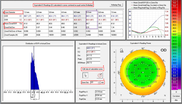
Methods: In this prospective comparative study, we included 100 myopic eyes of 50 patients with or without astigmatism. They were divided into two groups according to the platform used: WFO or WFG femtosecond LASIK. Using Holladay's equivalent keratometry reading (EKR) report of Pentacam HR, FOZ was defined as a zone centered on the pupil center with a standard deviation (SD) of 0.5 D, around the mean EKR. The differences in FOZ between the two platforms were analyzed at 3 months postoperatively. Visual acuity, refractive error, corneal asphericity (Q-value), and root mean square of higher-order aberrations (RMS for HOAs) were evaluated and compared.
Results: The mean (SD) of patient age was 26.64 (5.67) years. The preoperative characteristics of the two groups were comparable (all P > 0.05). The intended optical zone (IOZ) was 6 mm in both groups. The mean laser ablation depth was significantly greater in the WFG group (18 µm per D) than in the WFO group (16 µm per D) (P = 0.035). At 3 months postoperatively, the mean (SD) of FOZ diameter was 4.32 (0.94) mm (71.99% [15.68%] of intended optical zone) in the WFO group and 4.16 (1.13) mm (69.33% [18.78%] of intended optical zone) in the WFG group, with no significant difference between the two groups (P = 0.622). The change in corneal asphericity was greater in the WFG group than in the WFO group (P = 0.034). Postoperative mean corrected and uncorrected distance visual acuity, manifest refraction, and RMS for HOAs showed no significant difference between the two groups (all P > 0.05).
Conclusions: We found that WFG LASIK resulted in greater ablation depth and change in corneal asphericity than WFO LASIK at 3 months postoperatively. However, there was no significant difference in FOZ diameter, refractive error, and RMS for HOAs between the two groups. Further research is needed to confirm these findings.


 分享
分享
 求助内容:
求助内容: 应助结果提醒方式:
 扫码关注我们
