Optic nerve head perfusion changes in eyes with proliferative diabetic retinopathy treated with intravitreal ranibizumab or photocoagulation: a randomized controlled trial.
Ahmed Magdy Raffat Helmy, Mohammad Ahmad Rashad, Hesham Mohamed Gharieb, Wael Adel Gomaa, Rania Gamal Eldin Zaki
{"title":"Optic nerve head perfusion changes in eyes with proliferative diabetic retinopathy treated with intravitreal ranibizumab or photocoagulation: a randomized controlled trial.","authors":"Ahmed Magdy Raffat Helmy, Mohammad Ahmad Rashad, Hesham Mohamed Gharieb, Wael Adel Gomaa, Rania Gamal Eldin Zaki","doi":"10.51329/mehdiophthal1459","DOIUrl":null,"url":null,"abstract":"<p><strong>Background: </strong>Proliferative diabetic retinopathy (PDR) is a serious sight-threatening disease, and half of the patients with high-risk PDR can develop legal blindness within 5 years, if left untreated. This study was aimed at comparing panretinal photocoagulation (PRP) and intravitreal ranibizumab injections in terms of radial peripapillary capillary (RPC) density on optical coherence tomography angiography (OCTA) in patients with treatment-naive PDR.</p><p><strong>Methods: </strong>This open-label, prospective, randomized clinical trial included 50 patients with treatment-naive PDR with optic disc neovascularization and randomized them into two groups: group 1, with patients undergoing two sessions of PRP 2 weeks apart, and group 2, with patients received three intravitreal ranibizumab injections (0.5 mg) 1 month apart for 3 consecutive months. Patients underwent a full ophthalmological examination, including best-corrected distance visual acuity (BCDVA) measurement in the logarithm of minimal angle of resolution (logMAR) notation and OCTA before intervention and monthly after the last laser session or the first intravitreal ranibizumab injection for 3 months of follow-up. Visual field (VF) was tested at the beginning and end of 3 months.</p><p><strong>Results: </strong>Forty-two (84%) eyes completed the 3-month follow-up, including 22 eyes in the PRP group (88%) and 20 (80%) eyes in the ranibizumab group. The two groups were comparable in terms of demographic characteristics, diabetes duration, baseline BCDVA, glycated hemoglobin level, OCTA parameters, VF indices, and intraocular pressure (all <i>P</i> > 0.05). The RPC density change from baseline to the 3-month follow-up was significantly lower in the PRP group than in the ranibizumab group (mean difference in RPC density change: - 3.61%; 95% confidence interval: - 5.57% to - 1.60%; <i>P</i> = 0.001). The median (interquartile range) logMAR change from baseline to the 3-month follow-up (0.0 [0.2]) was significantly higher in the PRP group than in the ranibizumab group (- 0.15 [0.3]; <i>P</i> < 0.05). The median changes in central foveal thickness from baseline to the 3-month follow-up differed significantly between the two groups (<i>P</i> = 0.001).</p><p><strong>Conclusions: </strong>In eyes with PDR and neovascularization of the disc RPC density on OCTA increased in the ranibizumab group and decreased in the PRP group. Visual acuity gain was higher in the ranibizumab group than in the PRP group. Future multicenter trials addressing our limitations are required to verify the findings of this study.</p>","PeriodicalId":36524,"journal":{"name":"Medical Hypothesis, Discovery, and Innovation in Ophthalmology","volume":"11 4","pages":"144-150"},"PeriodicalIF":0.0000,"publicationDate":"2022-01-01","publicationTypes":"Journal Article","fieldsOfStudy":null,"isOpenAccess":false,"openAccessPdf":"https://ftp.ncbi.nlm.nih.gov/pub/pmc/oa_pdf/29/9b/mehdiophth-11-151.PMC10460245.pdf","citationCount":"0","resultStr":null,"platform":"Semanticscholar","paperid":null,"PeriodicalName":"Medical Hypothesis, Discovery, and Innovation in Ophthalmology","FirstCategoryId":"1085","ListUrlMain":"https://doi.org/10.51329/mehdiophthal1459","RegionNum":0,"RegionCategory":null,"ArticlePicture":[],"TitleCN":null,"AbstractTextCN":null,"PMCID":null,"EPubDate":"","PubModel":"","JCR":"Q2","JCRName":"Medicine","Score":null,"Total":0}
引用次数: 0
Abstract
Background: Proliferative diabetic retinopathy (PDR) is a serious sight-threatening disease, and half of the patients with high-risk PDR can develop legal blindness within 5 years, if left untreated. This study was aimed at comparing panretinal photocoagulation (PRP) and intravitreal ranibizumab injections in terms of radial peripapillary capillary (RPC) density on optical coherence tomography angiography (OCTA) in patients with treatment-naive PDR.
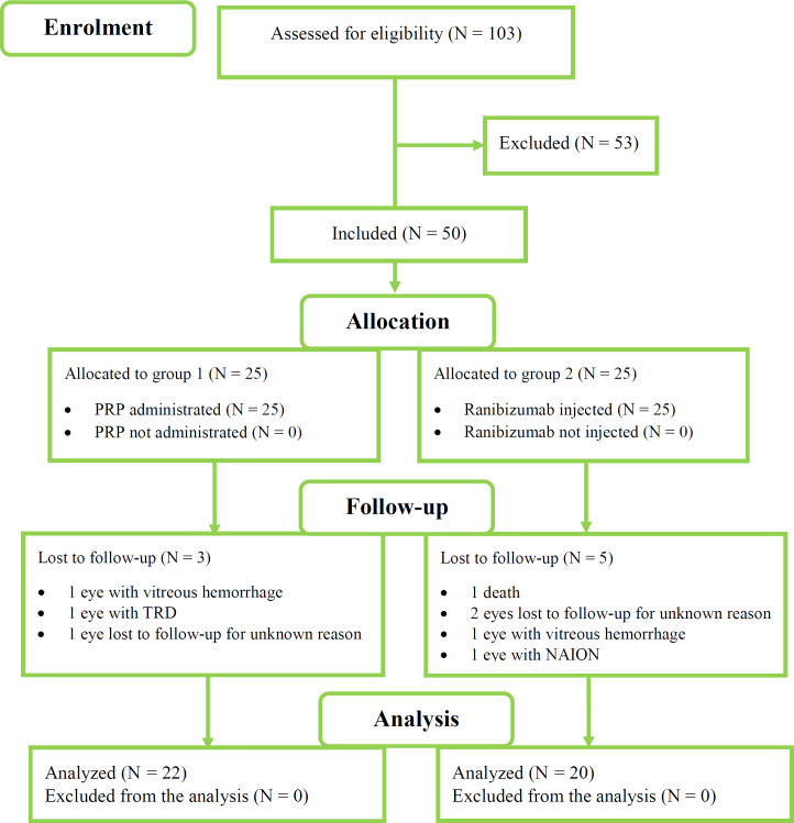
Methods: This open-label, prospective, randomized clinical trial included 50 patients with treatment-naive PDR with optic disc neovascularization and randomized them into two groups: group 1, with patients undergoing two sessions of PRP 2 weeks apart, and group 2, with patients received three intravitreal ranibizumab injections (0.5 mg) 1 month apart for 3 consecutive months. Patients underwent a full ophthalmological examination, including best-corrected distance visual acuity (BCDVA) measurement in the logarithm of minimal angle of resolution (logMAR) notation and OCTA before intervention and monthly after the last laser session or the first intravitreal ranibizumab injection for 3 months of follow-up. Visual field (VF) was tested at the beginning and end of 3 months.
Results: Forty-two (84%) eyes completed the 3-month follow-up, including 22 eyes in the PRP group (88%) and 20 (80%) eyes in the ranibizumab group. The two groups were comparable in terms of demographic characteristics, diabetes duration, baseline BCDVA, glycated hemoglobin level, OCTA parameters, VF indices, and intraocular pressure (all P > 0.05). The RPC density change from baseline to the 3-month follow-up was significantly lower in the PRP group than in the ranibizumab group (mean difference in RPC density change: - 3.61%; 95% confidence interval: - 5.57% to - 1.60%; P = 0.001). The median (interquartile range) logMAR change from baseline to the 3-month follow-up (0.0 [0.2]) was significantly higher in the PRP group than in the ranibizumab group (- 0.15 [0.3]; P < 0.05). The median changes in central foveal thickness from baseline to the 3-month follow-up differed significantly between the two groups (P = 0.001).
Conclusions: In eyes with PDR and neovascularization of the disc RPC density on OCTA increased in the ranibizumab group and decreased in the PRP group. Visual acuity gain was higher in the ranibizumab group than in the PRP group. Future multicenter trials addressing our limitations are required to verify the findings of this study.


 分享
分享
 求助内容:
求助内容: 应助结果提醒方式:
 扫码关注我们
