Jiangyan Zhao, Chenxing Ji, Haixia Cheng, Zhen Ye, Boyuan Yao, Ming Shen, Xuefei Shou, Xiang Zhou, Hongying Ye, Zhaoyun Zhang, Hong Chen, Yongfei Wang, Fuchu He, Yao Zhao, Wei Gong, Qilin Zhang, Nidan Qiao
{"title":"Digital image analysis allows objective stratification of patients with silent PIT1-lineage pituitary neuroendocrine tumors","authors":"Jiangyan Zhao, Chenxing Ji, Haixia Cheng, Zhen Ye, Boyuan Yao, Ming Shen, Xuefei Shou, Xiang Zhou, Hongying Ye, Zhaoyun Zhang, Hong Chen, Yongfei Wang, Fuchu He, Yao Zhao, Wei Gong, Qilin Zhang, Nidan Qiao","doi":"10.1002/cjp2.340","DOIUrl":null,"url":null,"abstract":"<p>Studies describing the clinical presentation and prognosis of patients with silent PIT1 (pituitary specific transcription factor)-lineage pituitary neuroendocrine tumors (PitNETs) are rare. We identified patients with positive PIT1 tumor staining but without evidence of hormone hypersecretion at a tertiary center. Clusters were obtained according to cell morphology and immunostaining from each patient's digitally segmented whole slide image. We compared the clinical presentations, radiological features, and prognoses of the different clusters. We identified 146 patients (68 male, 42.9 ± 14.1 years old) with silent PIT1-lineage PitNETs. Morphology clustering suggested that tumors with large nuclei and apparent eccentricity were associated with a higher proportion of aggressiveness and a higher hazard of recurrence [hazard ratio (HR): 2.64, (95% CI, 1.06–6.55), <i>p</i> = 0.037]. Immunohistochemical clustering suggested that tumors with thyroid stimulating hormone (TSH) staining or all negative PIT1-lineage hormones were associated with a higher proportion of aggressiveness and a higher risk of recurrence [HR: 12.4, (95% CI, 1.60–93.5), <i>p</i> = 0.015]. We obtained three-tier risk profiles by combining morphological and immunohistochemical clustering. Patients with the high-risk profile presented the highest recurrence rate compared with those in the medium-risk and low-risk profiles [HR: 3.54, (95% CI, 1.40–8.93), <i>p</i> = 0.002]. In conclusion, digital image analysis based on cell morphology and immunohistochemical staining allows objective stratification of patients with silent PIT1-lineage tumors. Typical morphological characteristics of high-risk tumors are large tumor nuclei and high eccentricity, and typical immunostaining characteristics are TSH staining or negative staining for all PIT1-lineage hormones.</p>","PeriodicalId":48612,"journal":{"name":"Journal of Pathology Clinical Research","volume":"9 6","pages":"488-497"},"PeriodicalIF":3.7000,"publicationDate":"2023-09-04","publicationTypes":"Journal Article","fieldsOfStudy":null,"isOpenAccess":false,"openAccessPdf":"https://pathsocjournals.onlinelibrary.wiley.com/doi/epdf/10.1002/cjp2.340","citationCount":"1","resultStr":null,"platform":"Semanticscholar","paperid":null,"PeriodicalName":"Journal of Pathology Clinical Research","FirstCategoryId":"3","ListUrlMain":"https://onlinelibrary.wiley.com/doi/10.1002/cjp2.340","RegionNum":2,"RegionCategory":"医学","ArticlePicture":[],"TitleCN":null,"AbstractTextCN":null,"PMCID":null,"EPubDate":"","PubModel":"","JCR":"Q1","JCRName":"PATHOLOGY","Score":null,"Total":0}
引用次数: 1
Abstract
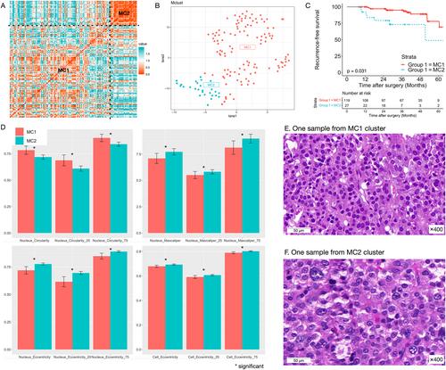
Studies describing the clinical presentation and prognosis of patients with silent PIT1 (pituitary specific transcription factor)-lineage pituitary neuroendocrine tumors (PitNETs) are rare. We identified patients with positive PIT1 tumor staining but without evidence of hormone hypersecretion at a tertiary center. Clusters were obtained according to cell morphology and immunostaining from each patient's digitally segmented whole slide image. We compared the clinical presentations, radiological features, and prognoses of the different clusters. We identified 146 patients (68 male, 42.9 ± 14.1 years old) with silent PIT1-lineage PitNETs. Morphology clustering suggested that tumors with large nuclei and apparent eccentricity were associated with a higher proportion of aggressiveness and a higher hazard of recurrence [hazard ratio (HR): 2.64, (95% CI, 1.06–6.55), p = 0.037]. Immunohistochemical clustering suggested that tumors with thyroid stimulating hormone (TSH) staining or all negative PIT1-lineage hormones were associated with a higher proportion of aggressiveness and a higher risk of recurrence [HR: 12.4, (95% CI, 1.60–93.5), p = 0.015]. We obtained three-tier risk profiles by combining morphological and immunohistochemical clustering. Patients with the high-risk profile presented the highest recurrence rate compared with those in the medium-risk and low-risk profiles [HR: 3.54, (95% CI, 1.40–8.93), p = 0.002]. In conclusion, digital image analysis based on cell morphology and immunohistochemical staining allows objective stratification of patients with silent PIT1-lineage tumors. Typical morphological characteristics of high-risk tumors are large tumor nuclei and high eccentricity, and typical immunostaining characteristics are TSH staining or negative staining for all PIT1-lineage hormones.
期刊介绍:
The Journal of Pathology: Clinical Research and The Journal of Pathology serve as translational bridges between basic biomedical science and clinical medicine with particular emphasis on, but not restricted to, tissue based studies.
The focus of The Journal of Pathology: Clinical Research is the publication of studies that illuminate the clinical relevance of research in the broad area of the study of disease. Appropriately powered and validated studies with novel diagnostic, prognostic and predictive significance, and biomarker discover and validation, will be welcomed. Studies with a predominantly mechanistic basis will be more appropriate for the companion Journal of Pathology.


 分享
分享
 求助内容:
求助内容: 应助结果提醒方式:
 扫码关注我们
