Filipe André Gonzalez, Miguel Ângelo-Dias, Catarina Martins, Rui Gomes, Jacobo Bacariza, Antero Fernandes, Luis Miguel Borrego
{"title":"Left atrial strain is associated with distinct inflammatory and immune profile in patients with COVID-19 pneumonia.","authors":"Filipe André Gonzalez, Miguel Ângelo-Dias, Catarina Martins, Rui Gomes, Jacobo Bacariza, Antero Fernandes, Luis Miguel Borrego","doi":"10.1186/s13089-022-00302-5","DOIUrl":null,"url":null,"abstract":"<p><strong>Introduction: </strong>SARS-CoV-2 infection is associated with multiple cardiac manifestations. Left atrial strain (LA-S) by speckle tracking echocardiography (STE) is a novel transthoracic echocardiography (TTE) measure of LA myocardial deformation and diastolic dysfunction, which could lead to early recognition of cardiac injury in severe COVID-19 patients with possible implications on clinical management, organ dysfunction, and mortality. Cardiac injury may occur by direct viral cytopathic effects or virus-driven immune activation, resulting in heart infiltration by inflammatory cells, despite limited and conflicting data are available on myocardial histology.</p><p><strong>Purpose: </strong>We aimed to explore LA-S and immune profiles in COVID-19 patients admitted to the intensive care unit (ICU) to identify distinctive features in patients with cardiac injury.</p><p><strong>Methods: </strong>We enrolled 30 patients > 18 years with positive SARS-CoV-2 RT-PCR, admitted to ICU. Acute myocardial infarction and pulmonary embolism were exclusion criteria. On days D1, D3, and D7 after ICU admission, patients performed TTE, hemogram, cardiac (pro-BNP; troponin) and inflammatory biomarkers (ESR; ferritin; IL1β; IL6; CRP; d-dimer; fibrinogen; PCT; adrenomedullin, ADM), and immunophenotyping by flow cytometry.</p><p><strong>Results: </strong>Patient's mean age was 60.7 y, with 63% males. Hypertension was the most common risk factor (73%; with 50% of patients under ACEi or ARA), followed by obesity (40%, mean BMI = 31 kg/m<sup>2</sup>). Cardiac dysfunction was detected by STE in 73% of patients: 40% left ventricle (LV) systolic dysfunction, 60% LV diastolic dysfunction, 37% right ventricle systolic dysfunction. Mortality, hospitalization days, remdesivir use, organ dysfunction, cardiac and serum biomarkers were not different between patients with (DYS) and without cardiac dysfunction (nDYS), except for ADM (increased in nDYS group at D7). From the 77 TTE, there was a striking difference between diastolic dysfunction evaluation by classic criteria compared to STE (28.6% vs. 57.1%, p = 0.0006). Lower reservoir (Ɛ) and contraction (ƐCT) LA-S correlated with IL-6 (Ɛ, p = 0.009, r = - 0.47; ƐCT, p = 0.0002, r = - 0.63) and central memory CD4 T-cells (ƐCT, p = 0.049, r = - 0.24). Along all timepoints, DYS patients showed persistent low lymphocyte counts that recovered at D7 in nDYS patients. DYS patients had lower platelets at D3 and showed a slower recovery in platelet counts and CRP levels; the latter significantly decreased at D7 in nDYS patients (p = 0.009). Overall, patients recovered with an increasing P/F ratio, though to a lesser extent in DYS patients.</p><p><strong>Discussion: </strong>Our study shows that LA-S may be a more sensitive marker for diastolic dysfunction in severe COVID-19, which could identify patients at risk for a protracted inflammatory state. A differential immune trait in DYS patients at ICU admission, with persistent lymphopenia, enriched CM T-cells, and higher IL-6 may suggest distinct inflammatory states or migration patterns in patients that develop cardiac injury.</p>","PeriodicalId":75201,"journal":{"name":"","volume":"15 1","pages":"2"},"PeriodicalIF":0.0,"publicationDate":"2023-01-12","publicationTypes":"Journal Article","fieldsOfStudy":null,"isOpenAccess":false,"openAccessPdf":"https://www.ncbi.nlm.nih.gov/pmc/articles/PMC9835022/pdf/","citationCount":"1","resultStr":null,"platform":"Semanticscholar","paperid":null,"PeriodicalName":"","FirstCategoryId":"1085","ListUrlMain":"https://doi.org/10.1186/s13089-022-00302-5","RegionNum":0,"RegionCategory":null,"ArticlePicture":[],"TitleCN":null,"AbstractTextCN":null,"PMCID":null,"EPubDate":"","PubModel":"","JCR":"","JCRName":"","Score":null,"Total":0}
引用次数: 1
Abstract
Introduction: SARS-CoV-2 infection is associated with multiple cardiac manifestations. Left atrial strain (LA-S) by speckle tracking echocardiography (STE) is a novel transthoracic echocardiography (TTE) measure of LA myocardial deformation and diastolic dysfunction, which could lead to early recognition of cardiac injury in severe COVID-19 patients with possible implications on clinical management, organ dysfunction, and mortality. Cardiac injury may occur by direct viral cytopathic effects or virus-driven immune activation, resulting in heart infiltration by inflammatory cells, despite limited and conflicting data are available on myocardial histology.
Purpose: We aimed to explore LA-S and immune profiles in COVID-19 patients admitted to the intensive care unit (ICU) to identify distinctive features in patients with cardiac injury.
Methods: We enrolled 30 patients > 18 years with positive SARS-CoV-2 RT-PCR, admitted to ICU. Acute myocardial infarction and pulmonary embolism were exclusion criteria. On days D1, D3, and D7 after ICU admission, patients performed TTE, hemogram, cardiac (pro-BNP; troponin) and inflammatory biomarkers (ESR; ferritin; IL1β; IL6; CRP; d-dimer; fibrinogen; PCT; adrenomedullin, ADM), and immunophenotyping by flow cytometry.
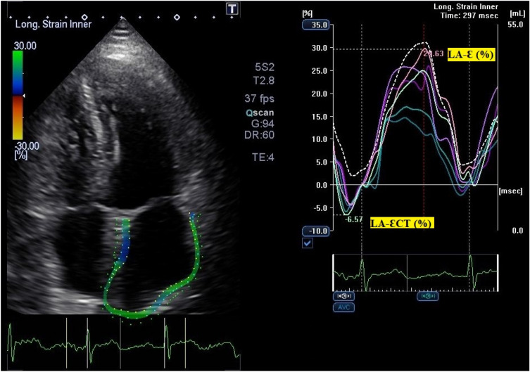
Results: Patient's mean age was 60.7 y, with 63% males. Hypertension was the most common risk factor (73%; with 50% of patients under ACEi or ARA), followed by obesity (40%, mean BMI = 31 kg/m2). Cardiac dysfunction was detected by STE in 73% of patients: 40% left ventricle (LV) systolic dysfunction, 60% LV diastolic dysfunction, 37% right ventricle systolic dysfunction. Mortality, hospitalization days, remdesivir use, organ dysfunction, cardiac and serum biomarkers were not different between patients with (DYS) and without cardiac dysfunction (nDYS), except for ADM (increased in nDYS group at D7). From the 77 TTE, there was a striking difference between diastolic dysfunction evaluation by classic criteria compared to STE (28.6% vs. 57.1%, p = 0.0006). Lower reservoir (Ɛ) and contraction (ƐCT) LA-S correlated with IL-6 (Ɛ, p = 0.009, r = - 0.47; ƐCT, p = 0.0002, r = - 0.63) and central memory CD4 T-cells (ƐCT, p = 0.049, r = - 0.24). Along all timepoints, DYS patients showed persistent low lymphocyte counts that recovered at D7 in nDYS patients. DYS patients had lower platelets at D3 and showed a slower recovery in platelet counts and CRP levels; the latter significantly decreased at D7 in nDYS patients (p = 0.009). Overall, patients recovered with an increasing P/F ratio, though to a lesser extent in DYS patients.
Discussion: Our study shows that LA-S may be a more sensitive marker for diastolic dysfunction in severe COVID-19, which could identify patients at risk for a protracted inflammatory state. A differential immune trait in DYS patients at ICU admission, with persistent lymphopenia, enriched CM T-cells, and higher IL-6 may suggest distinct inflammatory states or migration patterns in patients that develop cardiac injury.
简介:SARS-CoV-2感染与多种心脏表现相关。斑点跟踪超声心动图(STE)左心房应变(LA- s)是一种新型的经胸超声心动图(TTE)测量LA心肌变形和舒张功能障碍的方法,可以早期识别重症COVID-19患者的心脏损伤,可能对临床处理、器官功能障碍和死亡率有影响。心脏损伤可能由直接的病毒细胞病变作用或病毒驱动的免疫激活引起,导致炎症细胞浸润心脏,尽管心肌组织学数据有限且相互矛盾。目的:我们旨在探讨重症监护病房(ICU)入住的COVID-19患者的LA-S和免疫谱,以确定心脏损伤患者的独特特征。方法:选取30例> 18岁且SARS-CoV-2 RT-PCR阳性的ICU住院患者。急性心肌梗死和肺栓塞是排除标准。入院后第D1、D3、D7天,患者行TTE、血象、心肌(pro-BNP;肌钙蛋白)和炎症生物标志物(ESR;铁蛋白;摘要意思β;白细胞介素6;c反应蛋白;肺动脉栓塞;纤维蛋白原;PCT;肾上腺髓质素,ADM),并通过流式细胞术进行免疫分型。结果:患者平均年龄60.7岁,男性占63%。高血压是最常见的危险因素(73%;50%的患者处于ACEi或ARA状态),其次是肥胖(40%,平均BMI = 31 kg/m2)。73%的患者通过STE检测到心功能障碍:40%为左心室(LV)收缩功能障碍,60%为左心室舒张功能障碍,37%为右心室收缩功能障碍。死亡率、住院天数、瑞德西韦使用、器官功能障碍、心脏和血清生物标志物在患有(DYS)和没有心功能障碍(nDYS)的患者之间没有差异,除了ADM (nDYS组在D7时增加)。在77例TTE中,与STE相比,经典标准的舒张功能障碍评估有显著差异(28.6% vs. 57.1%, p = 0.0006)。下储层(Ɛ)和收缩(ƐCT) LA-S与IL-6相关(Ɛ, p = 0.009, r = - 0.47;ƐCT, p = 0.0002, r = - 0.63)和中央CD4记忆t细胞(ƐCT, p = 0.049, r = - 0.24)。在所有时间点上,DYS患者表现出持续的低淋巴细胞计数,nDYS患者在D7时恢复。DYS患者在D3时血小板较低,血小板计数和CRP水平恢复较慢;后者在nDYS患者的D7时显著降低(p = 0.009)。总体而言,患者的P/F比率增加,但DYS患者的P/F比率较低。讨论:我们的研究表明,LA-S可能是严重COVID-19患者舒张功能障碍的更敏感的标志物,可以识别有长期炎症状态风险的患者。在ICU入院的DYS患者中,持续淋巴细胞减少、CM t细胞丰富、IL-6升高等差异免疫特征可能表明,发生心脏损伤的患者存在不同的炎症状态或迁移模式。


 分享
分享
 求助内容:
求助内容: 应助结果提醒方式:
 扫码关注我们
