Amanda Lessa Martins, Anna Bárbara Scárdua Parreira, Maria Luiza Font Juliá Grossi, Raquel de Azevedo Benevides, Luciene Lage da Motta, Lucia Helena Sagrillo Pimassoni, Andrea Saade Daher Borjaili, Marcela Souza Lima Paulo, Danilo Nagib Salomão Paulo
{"title":"Study of the upper pole after subtotal splenectomy in rats.","authors":"Amanda Lessa Martins, Anna Bárbara Scárdua Parreira, Maria Luiza Font Juliá Grossi, Raquel de Azevedo Benevides, Luciene Lage da Motta, Lucia Helena Sagrillo Pimassoni, Andrea Saade Daher Borjaili, Marcela Souza Lima Paulo, Danilo Nagib Salomão Paulo","doi":"10.1590/acb371103","DOIUrl":null,"url":null,"abstract":"<p><strong>Purpose: </strong>To evaluate macro/microscopic viability of the upper pole (UP) in rats after 80 days of subtotal splenectomy preserving the upper pole (SSPUP).</p><p><strong>Methods: </strong>Twenty-five male Wistar rats were submitted to SSPUP. After 80 days, the rats were euthanized, and the remaining UP was evaluated macroscopically regarding appearance, color, consistency, length, width, thickness, and presence of fibrosis/necrosis; and microscopically regarding presence of red and white pulp, fibrosis/necrosis.</p><p><strong>Results: </strong>Two rats died during surgery and were removed from the statistical analysis. There was statistically significant increase in length and width between the pre and postoperative in the experimental group, with no significant difference in thickness. In the manipulation group, the macroscopic appearance of the spleen was normal in pre and postoperative, with viability preserved. In the experimental group, two UP of the spleen were not found during the second surgery. Macroscopically, it was observed absence of fibrosis and necrosis in all cases. Microscopically, the white and red pulp were intact in both groups. Two spleens of rats in the manipulation group presented areas with fibrosis and necrosis focus, which were not enough to be considered inviable.</p><p><strong>Conclusions: </strong>The UP of the spleen remained viable in 91.3% of the cases.</p>","PeriodicalId":6992,"journal":{"name":"Acta cirurgica brasileira","volume":"37 11","pages":"e371103"},"PeriodicalIF":1.3000,"publicationDate":"2023-01-01","publicationTypes":"Journal Article","fieldsOfStudy":null,"isOpenAccess":false,"openAccessPdf":"https://www.ncbi.nlm.nih.gov/pmc/articles/PMC9829190/pdf/","citationCount":"0","resultStr":null,"platform":"Semanticscholar","paperid":null,"PeriodicalName":"Acta cirurgica brasileira","FirstCategoryId":"3","ListUrlMain":"https://doi.org/10.1590/acb371103","RegionNum":4,"RegionCategory":"医学","ArticlePicture":[],"TitleCN":null,"AbstractTextCN":null,"PMCID":null,"EPubDate":"","PubModel":"","JCR":"Q3","JCRName":"SURGERY","Score":null,"Total":0}
引用次数: 0
Abstract
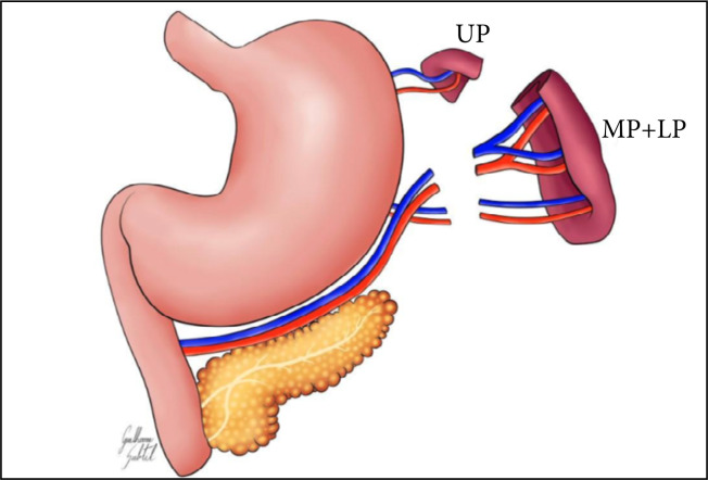
Purpose: To evaluate macro/microscopic viability of the upper pole (UP) in rats after 80 days of subtotal splenectomy preserving the upper pole (SSPUP).
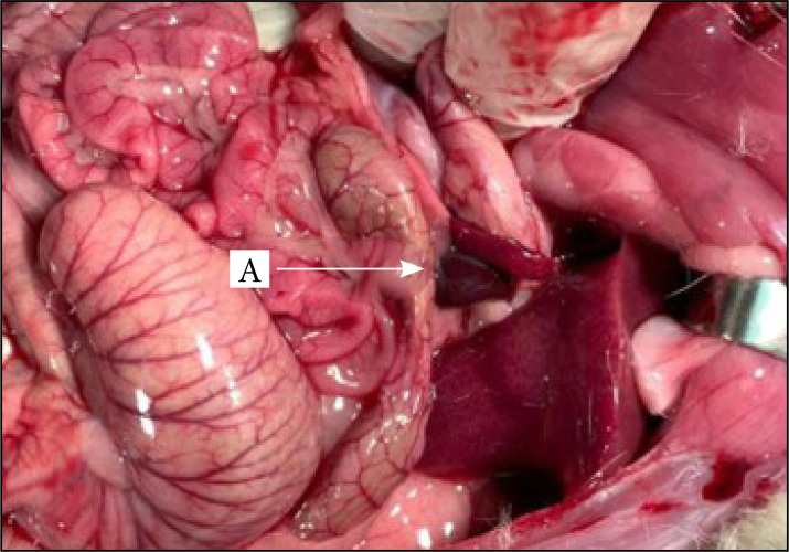
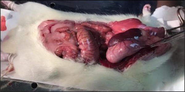
Methods: Twenty-five male Wistar rats were submitted to SSPUP. After 80 days, the rats were euthanized, and the remaining UP was evaluated macroscopically regarding appearance, color, consistency, length, width, thickness, and presence of fibrosis/necrosis; and microscopically regarding presence of red and white pulp, fibrosis/necrosis.
Results: Two rats died during surgery and were removed from the statistical analysis. There was statistically significant increase in length and width between the pre and postoperative in the experimental group, with no significant difference in thickness. In the manipulation group, the macroscopic appearance of the spleen was normal in pre and postoperative, with viability preserved. In the experimental group, two UP of the spleen were not found during the second surgery. Macroscopically, it was observed absence of fibrosis and necrosis in all cases. Microscopically, the white and red pulp were intact in both groups. Two spleens of rats in the manipulation group presented areas with fibrosis and necrosis focus, which were not enough to be considered inviable.
Conclusions: The UP of the spleen remained viable in 91.3% of the cases.


 分享
分享
 求助内容:
求助内容: 应助结果提醒方式:
 扫码关注我们
