Ahmet Özmeriç, Kadir Bahadır Alemdaroğlu, Ayşegül Fırat, Özgür Şahin
{"title":"Morphometric measurements for potential dangers of anterior intra-pelvic approach in women: A cadaveric study.","authors":"Ahmet Özmeriç, Kadir Bahadır Alemdaroğlu, Ayşegül Fırat, Özgür Şahin","doi":"10.5152/j.aott.2023.23013","DOIUrl":null,"url":null,"abstract":"<p><strong>Objective: </strong>This study aimed to improve the surgical anatomical knowledge of pelvic/acetabular trauma surgeons by providing detailed morphometric data on some of the most vulnerable arteries and nerves due to constant bony landmarks during anterior intra-pelvic approach fixation of acetabular fractures in women.</p><p><strong>Methods: </strong>Ten hemipelvis were dissected from 5 female cadavers. The following measurements relative to the symphysis were performed: (1) the distance of the corona mortis anastomosis and (2) the bisection of the external iliac vein with the pubic ramus. In addition, dis- tance to the pelvic brim at the level of pectineal convexity of the following structures was measured: (3) depth of obturatory neurovascu- lar bundle, (4) superior vesical artery, and (5) vaginal artery. Also, the clock position of the (6) gluteal superior and inferior vessels due to sciatic notch in the supine position. Due to antero-superior corner of sacroiliac joint (7) location of the common iliac artery bifurcation, (8) location of the bifurcation of internal iliac vessels to truncuses, (9) bifurcation of superior gluteal artery and lateral sacral artery, and (10) L5 nerve were measured. The descriptive statistics were given as medians and ranges as this is a descriptive anatomical study without comparisons.</p><p><strong>Results: </strong>The median distance of corona mortis to symphysis pubis was 59.5 mm (range = 58-61). The external iliac vein bisected the pubic arm 68.5 mm (range=65-70) lateral to the symphysis pubis. At the level of pectineal convexity (about the middle of the pelvic brim), obturatory neurovascular bundle, superior vesical artery, and vaginal artery were 15 mm (range=13-16), 24 mm (range=23-25), and 36 mm (range=34-38) inferior to the pelvic brim, respectively. The superior gluteal vessels leave the sciatic notch at 12 o'clock position in supine position. Inferior gluteal vessels leave the sciatic notch at 31⁄2 o'clock position (given for left side). Common iliac artery bifurcation bisects the SI joint 5 mm (4-7) superior to antero-superior corner of the Sacro-iliac (SI) joint. The internal iliac artery gives its posterior trunk 18 mm (range=15-20) straightly anterior to antero-superior corner of the SI joint. Bifurcation of superior gluteal artery and lateral sacral artery was 11 mm (range = 10-12) away from the beginning of the posterior truncus. L5 root's medial margin was 9 mm (range = 7-10) medial to this landmark, where its lateral margin was on the SI joint (2 mm medial to 2 mm lateral).</p><p><strong>Conclusion: </strong>The majority of the bleeding complications of the major branches of the internal and external iliac arteries and neurologic palsies due to obturatory nerve and L5 nerve root damage within the operative field of the anterior intra-pelvic approach can be avoided or managed by utilizing morphometric data provided from this study.</p><p><strong>Level of evidence: </strong>N/A.</p>","PeriodicalId":7097,"journal":{"name":"Acta orthopaedica et traumatologica turcica","volume":"57 4","pages":"183-188"},"PeriodicalIF":1.1000,"publicationDate":"2023-07-01","publicationTypes":"Journal Article","fieldsOfStudy":null,"isOpenAccess":false,"openAccessPdf":"https://ftp.ncbi.nlm.nih.gov/pub/pmc/oa_pdf/a7/b9/aott-57-4-183.PMC10544412.pdf","citationCount":"1","resultStr":null,"platform":"Semanticscholar","paperid":null,"PeriodicalName":"Acta orthopaedica et traumatologica turcica","FirstCategoryId":"3","ListUrlMain":"https://doi.org/10.5152/j.aott.2023.23013","RegionNum":4,"RegionCategory":"医学","ArticlePicture":[],"TitleCN":null,"AbstractTextCN":null,"PMCID":null,"EPubDate":"","PubModel":"","JCR":"Q3","JCRName":"ORTHOPEDICS","Score":null,"Total":0}
引用次数: 1
Abstract
Objective: This study aimed to improve the surgical anatomical knowledge of pelvic/acetabular trauma surgeons by providing detailed morphometric data on some of the most vulnerable arteries and nerves due to constant bony landmarks during anterior intra-pelvic approach fixation of acetabular fractures in women.
Methods: Ten hemipelvis were dissected from 5 female cadavers. The following measurements relative to the symphysis were performed: (1) the distance of the corona mortis anastomosis and (2) the bisection of the external iliac vein with the pubic ramus. In addition, dis- tance to the pelvic brim at the level of pectineal convexity of the following structures was measured: (3) depth of obturatory neurovascu- lar bundle, (4) superior vesical artery, and (5) vaginal artery. Also, the clock position of the (6) gluteal superior and inferior vessels due to sciatic notch in the supine position. Due to antero-superior corner of sacroiliac joint (7) location of the common iliac artery bifurcation, (8) location of the bifurcation of internal iliac vessels to truncuses, (9) bifurcation of superior gluteal artery and lateral sacral artery, and (10) L5 nerve were measured. The descriptive statistics were given as medians and ranges as this is a descriptive anatomical study without comparisons.
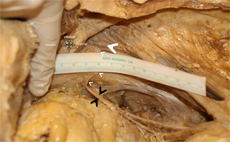
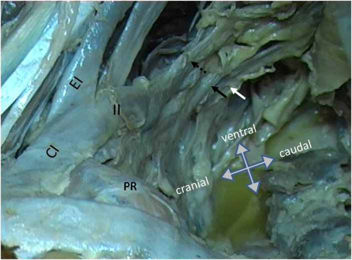
Results: The median distance of corona mortis to symphysis pubis was 59.5 mm (range = 58-61). The external iliac vein bisected the pubic arm 68.5 mm (range=65-70) lateral to the symphysis pubis. At the level of pectineal convexity (about the middle of the pelvic brim), obturatory neurovascular bundle, superior vesical artery, and vaginal artery were 15 mm (range=13-16), 24 mm (range=23-25), and 36 mm (range=34-38) inferior to the pelvic brim, respectively. The superior gluteal vessels leave the sciatic notch at 12 o'clock position in supine position. Inferior gluteal vessels leave the sciatic notch at 31⁄2 o'clock position (given for left side). Common iliac artery bifurcation bisects the SI joint 5 mm (4-7) superior to antero-superior corner of the Sacro-iliac (SI) joint. The internal iliac artery gives its posterior trunk 18 mm (range=15-20) straightly anterior to antero-superior corner of the SI joint. Bifurcation of superior gluteal artery and lateral sacral artery was 11 mm (range = 10-12) away from the beginning of the posterior truncus. L5 root's medial margin was 9 mm (range = 7-10) medial to this landmark, where its lateral margin was on the SI joint (2 mm medial to 2 mm lateral).
Conclusion: The majority of the bleeding complications of the major branches of the internal and external iliac arteries and neurologic palsies due to obturatory nerve and L5 nerve root damage within the operative field of the anterior intra-pelvic approach can be avoided or managed by utilizing morphometric data provided from this study.
期刊介绍:
Acta Orthopaedica et Traumatologica Turcica (AOTT) is an international, scientific, open access periodical published in accordance with independent, unbiased, and double-blinded peer-review principles. The journal is the official publication of the Turkish Association of Orthopaedics and Traumatology, and Turkish Society of Orthopaedics and Traumatology. It is published bimonthly in January, March, May, July, September, and November. The publication language of the journal is English.
The aim of the journal is to publish original studies of the highest scientific and clinical value in orthopedics, traumatology, and related disciplines. The scope of the journal includes but not limited to diagnostic, treatment, and prevention methods related to orthopedics and traumatology. Acta Orthopaedica et Traumatologica Turcica publishes clinical and basic research articles, case reports, personal clinical and technical notes, systematic reviews and meta-analyses and letters to the Editor. Proceedings of scientific meetings are also considered for publication.
The target audience of the journal includes healthcare professionals, physicians, and researchers who are interested or working in orthopedics and traumatology field, and related disciplines.


 分享
分享
 求助内容:
求助内容: 应助结果提醒方式:
 扫码关注我们
