Biodistribution of recombinant factor IX, extended half-life recombinant factor IX Fc fusion protein, and glycoPEGylated recombinant factor IX in hemophilia B mice.
Arjan van der Flier, Vu Hong, Zhan Liu, Peter Piepenhagen, Gregory Ulinski, Jennifer A Dumont, Kelly D Orcutt, Apollina Goel, Robert Peters, Joe Salas
{"title":"Biodistribution of recombinant factor IX, extended half-life recombinant factor IX Fc fusion protein, and glycoPEGylated recombinant factor IX in hemophilia B mice.","authors":"Arjan van der Flier, Vu Hong, Zhan Liu, Peter Piepenhagen, Gregory Ulinski, Jennifer A Dumont, Kelly D Orcutt, Apollina Goel, Robert Peters, Joe Salas","doi":"10.1097/MBC.0000000000001230","DOIUrl":null,"url":null,"abstract":"<p><p>Extended half-life recombinant FIX (rFIX) molecules have been generated to reduce the dosing burden and increase the protection of patients with hemophilia B. Clinical pharmacology studies with recombinant factor IX Fc fusion protein (rFIXFc) report a similar initial peak plasma recovery to that of rFIX, but with a larger volume of distribution. Although the pegylation of N9-GP results in a larger plasma recovery, there is a smaller volume of distribution, suggesting less extravasation of the latter drug. In this study, we set out to compare the biodistribution and tissue localization of rFIX, rFIXFc, and glycoPEGylated rFIX in a hemophilia B mouse model. Radiolabeled rFIX, rFIXFc, and rFIX-GP were employed in in vivo single-photon emission computed tomography imaging (SPECT/CT), microautoradiography (MARG), and histology to assess the distribution of FIX reagents over time. Immediately following injection, vascularized tissues demonstrated intense signal irrespective of FIX reagent. rFIX and rFIXFc were retained in joint and muscle areas through 5 half-lives, unlike rFIX-GP (assessed by SPECT). MARG and immunohistochemistry showed FIX agents localized at blood vessels among tissues, including liver, spleen, and kidney. Microautoradiographs, as well as fluorescent-labeled images of knee joint areas, demonstrated retention over time of FIX signal at the trabecular area of bone. Data indicate that rFIXFc is similar to rFIX in that it distributes outside the plasma compartment and is retained in certain tissues over time, while also retained at higher plasma levels. Overall, data suggest that Fc fusion does not impede the extravascular distribution of FIX.</p>","PeriodicalId":8992,"journal":{"name":"Blood Coagulation & Fibrinolysis","volume":"34 6","pages":"353-363"},"PeriodicalIF":1.1000,"publicationDate":"2023-09-01","publicationTypes":"Journal Article","fieldsOfStudy":null,"isOpenAccess":false,"openAccessPdf":"https://ftp.ncbi.nlm.nih.gov/pub/pmc/oa_pdf/fe/c9/blcof-34-353.PMC10481914.pdf","citationCount":"0","resultStr":null,"platform":"Semanticscholar","paperid":null,"PeriodicalName":"Blood Coagulation & Fibrinolysis","FirstCategoryId":"3","ListUrlMain":"https://doi.org/10.1097/MBC.0000000000001230","RegionNum":4,"RegionCategory":"医学","ArticlePicture":[],"TitleCN":null,"AbstractTextCN":null,"PMCID":null,"EPubDate":"2023/7/17 0:00:00","PubModel":"Epub","JCR":"Q4","JCRName":"HEMATOLOGY","Score":null,"Total":0}
引用次数: 0
Abstract
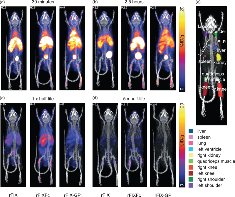
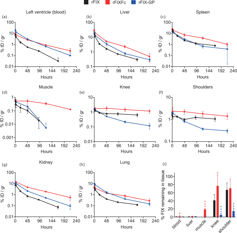
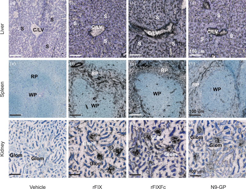
Extended half-life recombinant FIX (rFIX) molecules have been generated to reduce the dosing burden and increase the protection of patients with hemophilia B. Clinical pharmacology studies with recombinant factor IX Fc fusion protein (rFIXFc) report a similar initial peak plasma recovery to that of rFIX, but with a larger volume of distribution. Although the pegylation of N9-GP results in a larger plasma recovery, there is a smaller volume of distribution, suggesting less extravasation of the latter drug. In this study, we set out to compare the biodistribution and tissue localization of rFIX, rFIXFc, and glycoPEGylated rFIX in a hemophilia B mouse model. Radiolabeled rFIX, rFIXFc, and rFIX-GP were employed in in vivo single-photon emission computed tomography imaging (SPECT/CT), microautoradiography (MARG), and histology to assess the distribution of FIX reagents over time. Immediately following injection, vascularized tissues demonstrated intense signal irrespective of FIX reagent. rFIX and rFIXFc were retained in joint and muscle areas through 5 half-lives, unlike rFIX-GP (assessed by SPECT). MARG and immunohistochemistry showed FIX agents localized at blood vessels among tissues, including liver, spleen, and kidney. Microautoradiographs, as well as fluorescent-labeled images of knee joint areas, demonstrated retention over time of FIX signal at the trabecular area of bone. Data indicate that rFIXFc is similar to rFIX in that it distributes outside the plasma compartment and is retained in certain tissues over time, while also retained at higher plasma levels. Overall, data suggest that Fc fusion does not impede the extravascular distribution of FIX.
期刊介绍:
Blood Coagulation & Fibrinolysis is an international fully refereed journal that features review and original research articles on all clinical, laboratory and experimental aspects of haemostasis and thrombosis. The journal is devoted to publishing significant developments worldwide in the field of blood coagulation, fibrinolysis, thrombosis, platelets and the kininogen-kinin system, as well as dealing with those aspects of blood rheology relevant to haemostasis and the effects of drugs on haemostatic components


 分享
分享
 求助内容:
求助内容: 应助结果提醒方式:
 扫码关注我们
