{"title":"Posterior fossa giant adenoid cystic carcinoma with skull base invasion mimicking glomus jugulare: A case report and review of literature.","authors":"Anand Kumar Das, Saraj Kumar Singh, Kranti Bhavana, Subhash Kumar","doi":"10.1177/20363613221150218","DOIUrl":null,"url":null,"abstract":"<p><p>The author describes a rare case of giant adenoid cystic carcinoma (ACC) mimicking large paraganglioma with lower cranial nerve palsy. A 60-year-old female presented with a progressive increase in postauricular swelling with unilateral hearing loss, facial deviation, difficulty in swallowing, and hoarseness of voice. MRI brain showed highly vascular infiltrating and osteolytic mass suggestive of large glomus jugulare versus sarcoma. It was completely engulfing the jugular foramen and lower cranial nerves with bony erosion of the jugular foramen and occipital condyle. The whole mastoid was filled with the tumor. On digital subtraction angiography the majority of blood supply was from the occipital branch of the external carotid artery and vertebral artery. The patient underwent percutaneous embolization followed by external carotid ligation and resection of the mass. The postoperative course was uneventful. Histopathology was suggestive of mixed ACCs. The patient received radiotherapy. After 1 year of follow up no recurrence or distant metastasis was noted.</p>","PeriodicalId":46078,"journal":{"name":"Rare Tumors","volume":"15 ","pages":"20363613221150218"},"PeriodicalIF":0.9000,"publicationDate":"2023-01-01","publicationTypes":"Journal Article","fieldsOfStudy":null,"isOpenAccess":false,"openAccessPdf":"https://ftp.ncbi.nlm.nih.gov/pub/pmc/oa_pdf/db/e8/10.1177_20363613221150218.PMC9830093.pdf","citationCount":"0","resultStr":null,"platform":"Semanticscholar","paperid":null,"PeriodicalName":"Rare Tumors","FirstCategoryId":"1085","ListUrlMain":"https://doi.org/10.1177/20363613221150218","RegionNum":0,"RegionCategory":null,"ArticlePicture":[],"TitleCN":null,"AbstractTextCN":null,"PMCID":null,"EPubDate":"","PubModel":"","JCR":"Q4","JCRName":"ONCOLOGY","Score":null,"Total":0}
引用次数: 0
Abstract
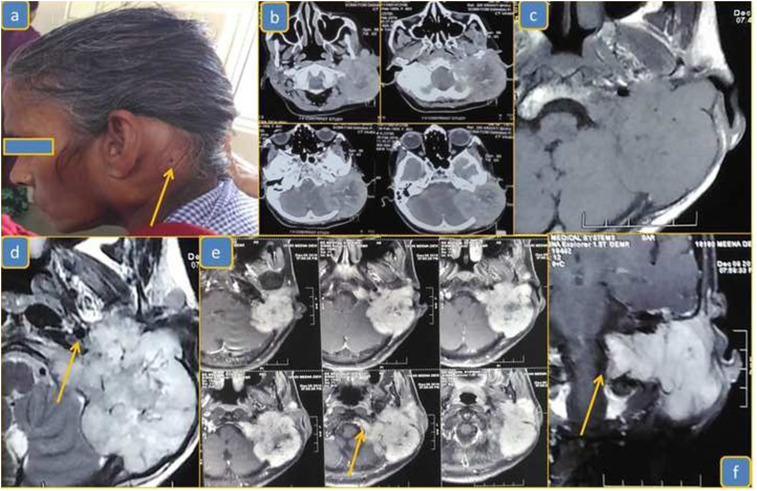
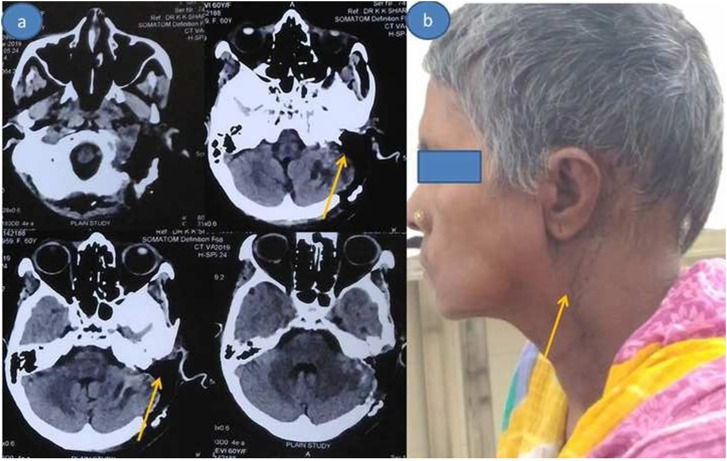
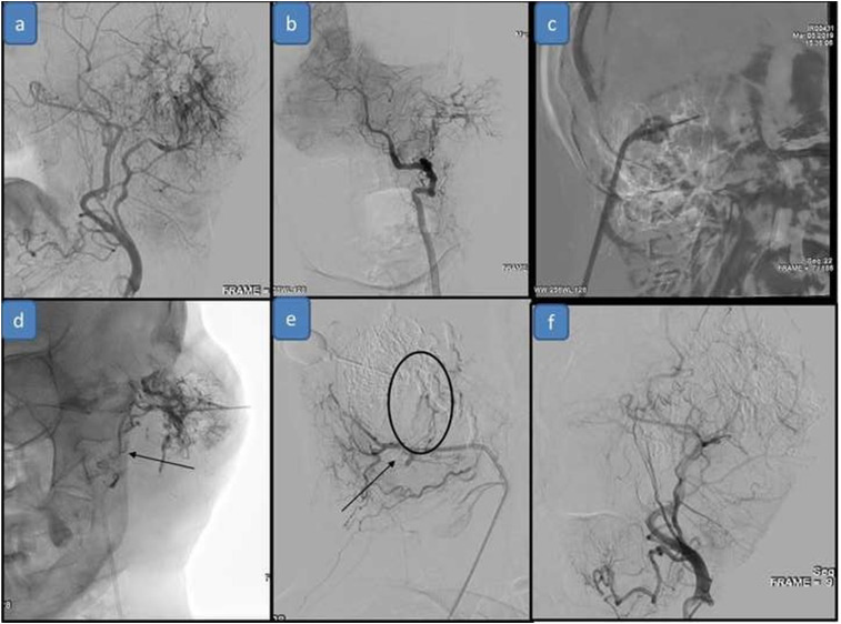
The author describes a rare case of giant adenoid cystic carcinoma (ACC) mimicking large paraganglioma with lower cranial nerve palsy. A 60-year-old female presented with a progressive increase in postauricular swelling with unilateral hearing loss, facial deviation, difficulty in swallowing, and hoarseness of voice. MRI brain showed highly vascular infiltrating and osteolytic mass suggestive of large glomus jugulare versus sarcoma. It was completely engulfing the jugular foramen and lower cranial nerves with bony erosion of the jugular foramen and occipital condyle. The whole mastoid was filled with the tumor. On digital subtraction angiography the majority of blood supply was from the occipital branch of the external carotid artery and vertebral artery. The patient underwent percutaneous embolization followed by external carotid ligation and resection of the mass. The postoperative course was uneventful. Histopathology was suggestive of mixed ACCs. The patient received radiotherapy. After 1 year of follow up no recurrence or distant metastasis was noted.


 分享
分享
 求助内容:
求助内容: 应助结果提醒方式:
 扫码关注我们
