Chao Lin , Li Zhang , Ziying Zhang , Yifeng Jiang , Xueming Li
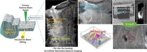
{"title":"Locating cellular contents during cryoFIB milling using cellular secondary-electron imaging","authors":"Chao Lin , Li Zhang , Ziying Zhang , Yifeng Jiang , Xueming Li","doi":"10.1016/j.jsb.2023.108005","DOIUrl":null,"url":null,"abstract":"<div><p>Cryo-electron tomography (cryoET) is a powerful technology that allows <em>in-situ</em> observation of the molecular structure of tissues and cells. Cryo-focused ion beam (cryoFIB) milling plays an important role in the preparation of high-quality thin lamellar samples for cryoET studies, thus, promoting the rapid development of cryoET in recent years. However, locating the regions of interest in a large cell or tissue during cryoFIB milling remains a major challenge limiting cryoET applications on arbitrary biological samples. Here, we report an on-the-fly localization method based on cellular secondary electron imaging (CSEI), which is derived from a basic imaging function of the cryoFIB instruments and enables high-contrast imaging of the cellular contents of frozen-hydrated biological samples. Moreover, CSEI does not require fluorescent labels and additional devices. The present study discusses the imaging principles and settings for optimizing CSEI. Tests on several commercially available cryoFIB instruments demonstrated that CSEI was feasible on mainstream instruments to observe all types of cellular contents and reliable under different milling conditions. We established a simple milling-localization workflow and tested it using the basal body of <em>Chlamydomonas reinhardtii</em>.</p></div>","PeriodicalId":17074,"journal":{"name":"Journal of structural biology","volume":"215 3","pages":"Article 108005"},"PeriodicalIF":2.7000,"publicationDate":"2023-09-01","publicationTypes":"Journal Article","fieldsOfStudy":null,"isOpenAccess":false,"openAccessPdf":"","citationCount":"0","resultStr":null,"platform":"Semanticscholar","paperid":null,"PeriodicalName":"Journal of structural biology","FirstCategoryId":"99","ListUrlMain":"https://www.sciencedirect.com/science/article/pii/S1047847723000680","RegionNum":3,"RegionCategory":"生物学","ArticlePicture":[],"TitleCN":null,"AbstractTextCN":null,"PMCID":null,"EPubDate":"","PubModel":"","JCR":"Q3","JCRName":"BIOCHEMISTRY & MOLECULAR BIOLOGY","Score":null,"Total":0}
引用次数: 0
Abstract
Cryo-electron tomography (cryoET) is a powerful technology that allows in-situ observation of the molecular structure of tissues and cells. Cryo-focused ion beam (cryoFIB) milling plays an important role in the preparation of high-quality thin lamellar samples for cryoET studies, thus, promoting the rapid development of cryoET in recent years. However, locating the regions of interest in a large cell or tissue during cryoFIB milling remains a major challenge limiting cryoET applications on arbitrary biological samples. Here, we report an on-the-fly localization method based on cellular secondary electron imaging (CSEI), which is derived from a basic imaging function of the cryoFIB instruments and enables high-contrast imaging of the cellular contents of frozen-hydrated biological samples. Moreover, CSEI does not require fluorescent labels and additional devices. The present study discusses the imaging principles and settings for optimizing CSEI. Tests on several commercially available cryoFIB instruments demonstrated that CSEI was feasible on mainstream instruments to observe all types of cellular contents and reliable under different milling conditions. We established a simple milling-localization workflow and tested it using the basal body of Chlamydomonas reinhardtii.
期刊介绍:
Journal of Structural Biology (JSB) has an open access mirror journal, the Journal of Structural Biology: X (JSBX), sharing the same aims and scope, editorial team, submission system and rigorous peer review. Since both journals share the same editorial system, you may submit your manuscript via either journal homepage. You will be prompted during submission (and revision) to choose in which to publish your article. The editors and reviewers are not aware of the choice you made until the article has been published online. JSB and JSBX publish papers dealing with the structural analysis of living material at every level of organization by all methods that lead to an understanding of biological function in terms of molecular and supermolecular structure.
Techniques covered include:
• Light microscopy including confocal microscopy
• All types of electron microscopy
• X-ray diffraction
• Nuclear magnetic resonance
• Scanning force microscopy, scanning probe microscopy, and tunneling microscopy
• Digital image processing
• Computational insights into structure


 分享
分享
 求助内容:
求助内容: 应助结果提醒方式:
 扫码关注我们
