{"title":"[Three-dimensional analysis of posttraumatic tibial shaft malunion and correction based on the healthy, contralateral leg].","authors":"Arnd F Viehöfer, Stephan H Wirth","doi":"10.1007/s00064-023-00821-x","DOIUrl":null,"url":null,"abstract":"<p><strong>Objective: </strong>Three-dimensional (3D) analysis and implementation with patient-specific cutting and repositioning blocks enables correction of complex tibial malunions. Correction can be planned using the contralateral side or a statistical model. Patient-specific 3D-printed cutting guide blocks enable a precise osteotomy and reduction guide blocks help to achieve anatomical reduction. Depending on the type and extent of correction, fibula osteotomy may need to be considered to achieve the desired reduction.</p><p><strong>Contraindications: </strong>a) Poor soft tissue (flap surgery, adherent skin in field of operation); b) infection; c) peripheral artery disease (stage III and IV classified according to Fontaine, critical transcutaneous oxygen partial pressure, TcPO<sub>2</sub>); d) general contraindication to surgery.</p><p><strong>Surgical technique: </strong>Before surgery, a 3D model of both lower legs is created based on computed tomography (CT) scans. Analysis of the deformity based on the contralateral side in a 3D computer model (CASPA) and planning of the osteotomy. If the contralateral side also has a deformity, a statistical model can be used. Printing of patient-specific guides made of nylon (PA2200) for the osteotomy and reduction. Surgery is performed in supine position, antibiotic prophylaxis, thigh tourniquet, which is used as needed. Ventrolateral approach to the tibia. Attachment of the patient-specific osteotomy guide, performance of the osteotomy. Reduction using the guide. Fibula osteotomy through a lateral approach is performed if the reduction of the tibia is hindered by the fibula. This can be performed freehand or with patient-specific guides. Wound closure.</p><p><strong>Postoperative management: </strong>Compartment monitoring. Passive mobilization of the ankle in the cast as soon as the wound healing has progressed. Partial weightbearing in a lower leg cast for at least 6-12 weeks, depending on the routinely performed radiographic assessment 6 weeks postoperatively. Thromboprophylaxis with low molecular weight heparin until cast removal.</p><p><strong>Results: </strong>Patient-specific correction of malunions are generally good. This could be confirmed for distal tibial corrections. For tibial shaft deformities, the final results are still pending. Preliminary results, however, show good feasibility with a pseudarthrosis rate of 10% without postoperative infection.</p>","PeriodicalId":54677,"journal":{"name":"Operative Orthopadie Und Traumatologie","volume":" ","pages":"239-247"},"PeriodicalIF":1.0000,"publicationDate":"2023-10-01","publicationTypes":"Journal Article","fieldsOfStudy":null,"isOpenAccess":false,"openAccessPdf":"https://www.ncbi.nlm.nih.gov/pmc/articles/PMC10520191/pdf/","citationCount":"0","resultStr":null,"platform":"Semanticscholar","paperid":null,"PeriodicalName":"Operative Orthopadie Und Traumatologie","FirstCategoryId":"3","ListUrlMain":"https://doi.org/10.1007/s00064-023-00821-x","RegionNum":4,"RegionCategory":"医学","ArticlePicture":[],"TitleCN":null,"AbstractTextCN":null,"PMCID":null,"EPubDate":"2023/9/12 0:00:00","PubModel":"Epub","JCR":"Q3","JCRName":"ORTHOPEDICS","Score":null,"Total":0}
引用次数: 0
Abstract
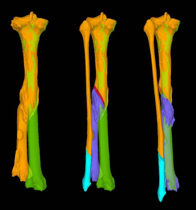
Objective: Three-dimensional (3D) analysis and implementation with patient-specific cutting and repositioning blocks enables correction of complex tibial malunions. Correction can be planned using the contralateral side or a statistical model. Patient-specific 3D-printed cutting guide blocks enable a precise osteotomy and reduction guide blocks help to achieve anatomical reduction. Depending on the type and extent of correction, fibula osteotomy may need to be considered to achieve the desired reduction.
Contraindications: a) Poor soft tissue (flap surgery, adherent skin in field of operation); b) infection; c) peripheral artery disease (stage III and IV classified according to Fontaine, critical transcutaneous oxygen partial pressure, TcPO2); d) general contraindication to surgery.
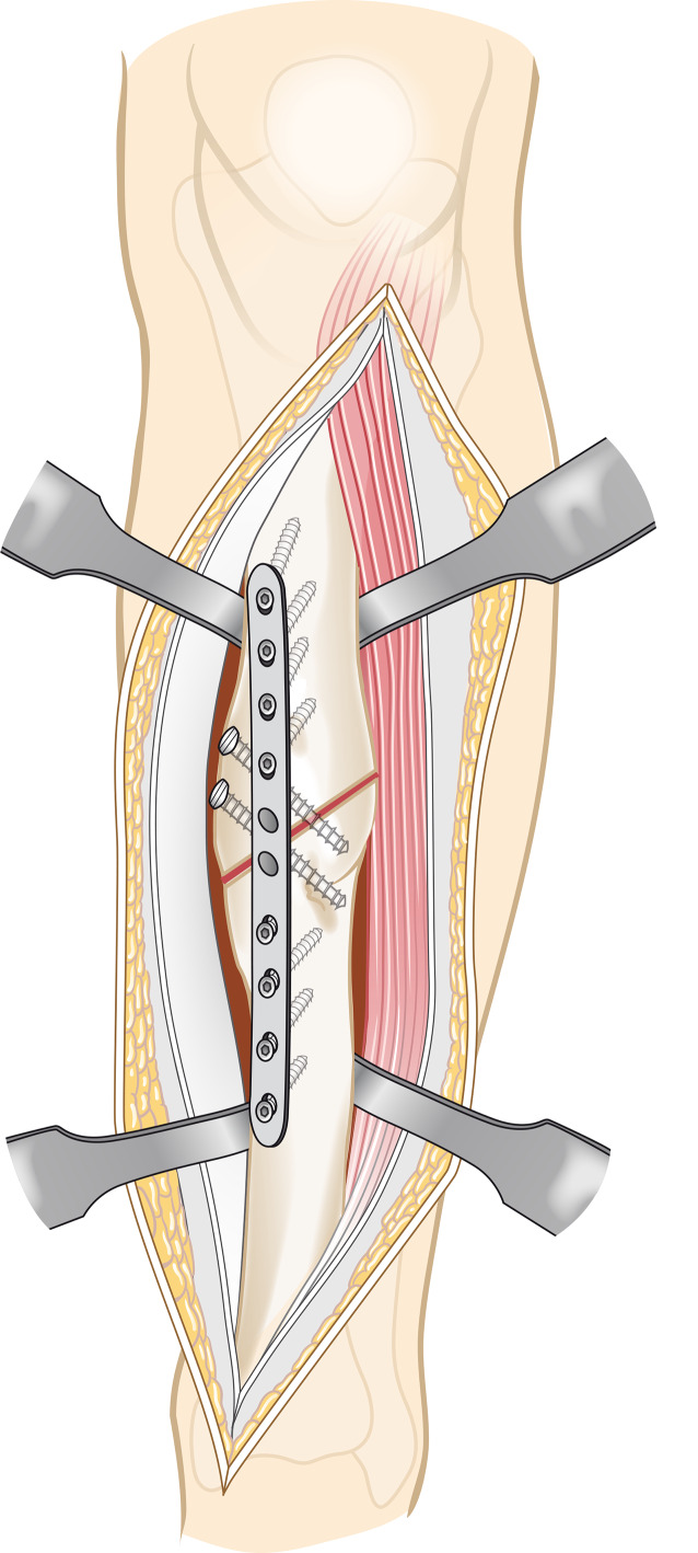
Surgical technique: Before surgery, a 3D model of both lower legs is created based on computed tomography (CT) scans. Analysis of the deformity based on the contralateral side in a 3D computer model (CASPA) and planning of the osteotomy. If the contralateral side also has a deformity, a statistical model can be used. Printing of patient-specific guides made of nylon (PA2200) for the osteotomy and reduction. Surgery is performed in supine position, antibiotic prophylaxis, thigh tourniquet, which is used as needed. Ventrolateral approach to the tibia. Attachment of the patient-specific osteotomy guide, performance of the osteotomy. Reduction using the guide. Fibula osteotomy through a lateral approach is performed if the reduction of the tibia is hindered by the fibula. This can be performed freehand or with patient-specific guides. Wound closure.
Postoperative management: Compartment monitoring. Passive mobilization of the ankle in the cast as soon as the wound healing has progressed. Partial weightbearing in a lower leg cast for at least 6-12 weeks, depending on the routinely performed radiographic assessment 6 weeks postoperatively. Thromboprophylaxis with low molecular weight heparin until cast removal.
Results: Patient-specific correction of malunions are generally good. This could be confirmed for distal tibial corrections. For tibial shaft deformities, the final results are still pending. Preliminary results, however, show good feasibility with a pseudarthrosis rate of 10% without postoperative infection.
期刊介绍:
Orthopedics and Traumatology is directed toward all orthopedic surgeons, trauma-tologists, hand surgeons, specialists in sports injuries, orthopedics and rheumatology as well as gene-al surgeons who require access to reliable information on current operative methods to ensure the quality of patient advice, preoperative planning, and postoperative care.
The journal presents established and new operative procedures in uniformly structured and extensively illustrated contributions. All aspects are presented step-by-step from indications, contraindications, patient education, and preparation of the operation right through to postoperative care. The advantages and disadvantages, possible complications, deficiencies and risks of the methods as well as significant results with their evaluation criteria are discussed. To allow the reader to assess the outcome, results are detailed and based on internationally recognized scoring systems.
Orthopedics and Traumatology facilitates effective advancement and further education for all those active in both special and conservative fields of orthopedics, traumatology, and general surgery, offers sup-port for therapeutic decision-making, and provides – more than 30 years after its first publication – constantly expanding and up-to-date teaching on operative techniques.


 分享
分享
 求助内容:
求助内容: 应助结果提醒方式:
 扫码关注我们
