{"title":"Two cases of cartilaginous metaplasia in the sclera of Japanese White rabbits.","authors":"Kotaro Yamada, Yoshinori Yamagiwa, Miki Masatsugu, Yu Haranosono","doi":"10.1293/tox.2022-0062","DOIUrl":null,"url":null,"abstract":"<p><p>Spontaneous cartilaginous metaplasia of the sclera has not been reported in rabbits. Herein, we report two cases of spontaneous cartilaginous metaplasia in the sclera of Japanese White (JW) rabbits. Case 1 was noted in a 14-week-old male Kbs:JW rabbit that received a single ocular instillation of 20% isoproterenol (IP) a day before necropsy, and showed no abnormalities in clinical signs, ophthalmological assessments, and necropsy. Case 2 was noted in a 38-week-old male Kbs:JW rabbit that was housed under light-emitting diode (LED) lighting for 26 weeks and showed no effects of LED on clinical signs, ophthalmological assessments, and necropsy. Histological sections of the eyes of both animals were prepared and stained with hematoxylin and eosin (H&E) and Alcian blue, and immunohistochemical staining for vimentin was performed. The H&E-stained specimens showed focal hyaline cartilage-like tissues distributed between the scleral fibers at the posterior pole in both cases. The surrounding scleral fibers were compressed and/or partially destroyed by the cartilage-like tissue. The cartilage-like matrix was stained blue by Alcian blue, and immunohistochemistry showed that chondrocyte-like cells were positive for vimentin. Based on these findings, we diagnosed cartilaginous metaplasia in the sclera of Kbs:JW rabbits. The lesion was farther from the IP administration site in Case 1 and was not accompanied by other ophthalmological or histopathological abnormalities in either of the cases. This implies that the lesions occurred spontaneously owing to the abnormal differentiation of neural crest-derived cells.</p>","PeriodicalId":17437,"journal":{"name":"Journal of Toxicologic Pathology","volume":"36 1","pages":"45-48"},"PeriodicalIF":0.9000,"publicationDate":"2023-01-01","publicationTypes":"Journal Article","fieldsOfStudy":null,"isOpenAccess":false,"openAccessPdf":"https://ftp.ncbi.nlm.nih.gov/pub/pmc/oa_pdf/f0/de/tox-36-045.PMC9837471.pdf","citationCount":"0","resultStr":null,"platform":"Semanticscholar","paperid":null,"PeriodicalName":"Journal of Toxicologic Pathology","FirstCategoryId":"3","ListUrlMain":"https://doi.org/10.1293/tox.2022-0062","RegionNum":4,"RegionCategory":"医学","ArticlePicture":[],"TitleCN":null,"AbstractTextCN":null,"PMCID":null,"EPubDate":"","PubModel":"","JCR":"Q4","JCRName":"PATHOLOGY","Score":null,"Total":0}
引用次数: 0
Abstract
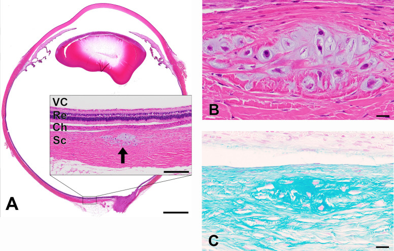
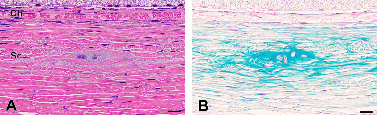
Spontaneous cartilaginous metaplasia of the sclera has not been reported in rabbits. Herein, we report two cases of spontaneous cartilaginous metaplasia in the sclera of Japanese White (JW) rabbits. Case 1 was noted in a 14-week-old male Kbs:JW rabbit that received a single ocular instillation of 20% isoproterenol (IP) a day before necropsy, and showed no abnormalities in clinical signs, ophthalmological assessments, and necropsy. Case 2 was noted in a 38-week-old male Kbs:JW rabbit that was housed under light-emitting diode (LED) lighting for 26 weeks and showed no effects of LED on clinical signs, ophthalmological assessments, and necropsy. Histological sections of the eyes of both animals were prepared and stained with hematoxylin and eosin (H&E) and Alcian blue, and immunohistochemical staining for vimentin was performed. The H&E-stained specimens showed focal hyaline cartilage-like tissues distributed between the scleral fibers at the posterior pole in both cases. The surrounding scleral fibers were compressed and/or partially destroyed by the cartilage-like tissue. The cartilage-like matrix was stained blue by Alcian blue, and immunohistochemistry showed that chondrocyte-like cells were positive for vimentin. Based on these findings, we diagnosed cartilaginous metaplasia in the sclera of Kbs:JW rabbits. The lesion was farther from the IP administration site in Case 1 and was not accompanied by other ophthalmological or histopathological abnormalities in either of the cases. This implies that the lesions occurred spontaneously owing to the abnormal differentiation of neural crest-derived cells.
期刊介绍:
JTP is a scientific journal that publishes original studies in the field of toxicological pathology and in a wide variety of other related fields. The main scope of the journal is listed below.
Administrative Opinions of Policymakers and Regulatory Agencies
Adverse Events
Carcinogenesis
Data of A Predominantly Negative Nature
Drug-Induced Hematologic Toxicity
Embryological Pathology
High Throughput Pathology
Historical Data of Experimental Animals
Immunohistochemical Analysis
Molecular Pathology
Nomenclature of Lesions
Non-mammal Toxicity Study
Result or Lesion Induced by Chemicals of Which Names Hidden on Account of the Authors
Technology and Methodology Related to Toxicological Pathology
Tumor Pathology; Neoplasia and Hyperplasia
Ultrastructural Analysis
Use of Animal Models.


 分享
分享
 求助内容:
求助内容: 应助结果提醒方式:
 扫码关注我们
