{"title":"The Role of Endoplasmic Reticulum Stress in Cell Injury Induced by Methimazole on Pancreatic Cells.","authors":"Özge Yazıcı, Mehtap Kara, Tuğçe Boran, Gul Ozhan","doi":"10.34172/apb.2023.042","DOIUrl":null,"url":null,"abstract":"Purpose: Methimazole is an anti-thyroid agent, especially as main therapy option for Graves’ disease in children and adults. Drug induced pancreatitis is one of the known adverse effect of methimazole mentioned in case reports. However, the detailed molecular mechanisms of methimazole-induced pancreatitis are still unclear. In this study, the aim is to investigate the adverse effect of methimazole on pancreas cell stress mechanism and apoptosis. Methods: Cytotoxicity was evaluated in human pancreas/duct (PANC-1) cell line. Total oxidant (TOS) and antioxidant status (TAS) for oxidative stress index, glutathione (GSH) level and endoplasmic reticulum (ER) stress biomarkers were evaluated by ELISA. Reactive oxygen species (ROS) levels and apoptosis were evaluated by flow-cytometer. Results: The 30% inhibition rate concentration (IC30) value was determined as 53 mM in PANC1 cells. The exposure concentrations were in the range of 0-40 mM for 48 hours. Methimazole might induce cellular stress conditions. ROS production increases depending on concentration, and this increase shows parallelism with the increase in ER stress biomarkers such as TOS, ERN1 and CASPASE12. Conversely, there was no significant difference between control and exposure groups in terms of apoptosis. Conclusion: In conclusion, methimazole might have triggered the mechanisms of inflammation or autophagy in the pancreatic cells. However, there is still a need for in vitro and in vivo studies including other cellular parameters related to apoptosis.","PeriodicalId":7256,"journal":{"name":"Advanced pharmaceutical bulletin","volume":"13 1","pages":"196-201"},"PeriodicalIF":4.1000,"publicationDate":"2023-01-01","publicationTypes":"Journal Article","fieldsOfStudy":null,"isOpenAccess":false,"openAccessPdf":"https://www.ncbi.nlm.nih.gov/pmc/articles/PMC9871271/pdf/","citationCount":"0","resultStr":null,"platform":"Semanticscholar","paperid":null,"PeriodicalName":"Advanced pharmaceutical bulletin","FirstCategoryId":"1085","ListUrlMain":"https://doi.org/10.34172/apb.2023.042","RegionNum":0,"RegionCategory":null,"ArticlePicture":[],"TitleCN":null,"AbstractTextCN":null,"PMCID":null,"EPubDate":"","PubModel":"","JCR":"Q2","JCRName":"PHARMACOLOGY & PHARMACY","Score":null,"Total":0}
引用次数: 0
Abstract
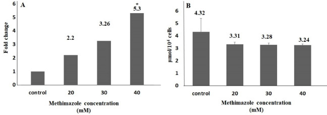
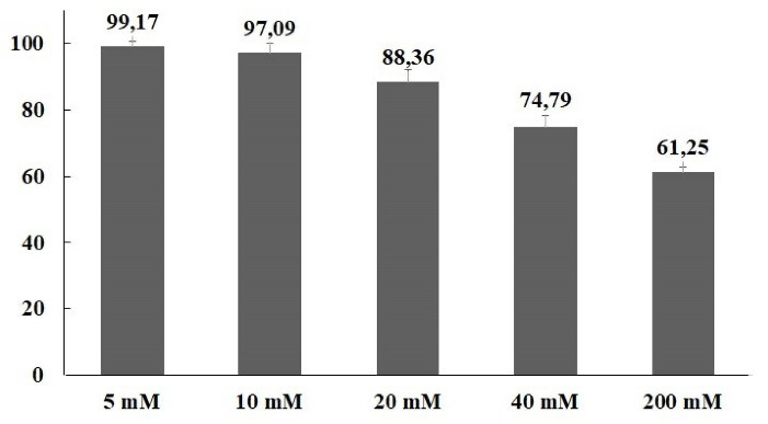
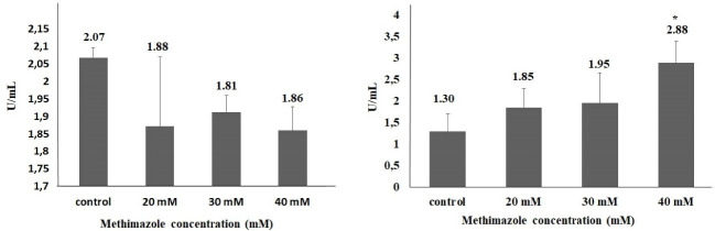
Purpose: Methimazole is an anti-thyroid agent, especially as main therapy option for Graves’ disease in children and adults. Drug induced pancreatitis is one of the known adverse effect of methimazole mentioned in case reports. However, the detailed molecular mechanisms of methimazole-induced pancreatitis are still unclear. In this study, the aim is to investigate the adverse effect of methimazole on pancreas cell stress mechanism and apoptosis. Methods: Cytotoxicity was evaluated in human pancreas/duct (PANC-1) cell line. Total oxidant (TOS) and antioxidant status (TAS) for oxidative stress index, glutathione (GSH) level and endoplasmic reticulum (ER) stress biomarkers were evaluated by ELISA. Reactive oxygen species (ROS) levels and apoptosis were evaluated by flow-cytometer. Results: The 30% inhibition rate concentration (IC30) value was determined as 53 mM in PANC1 cells. The exposure concentrations were in the range of 0-40 mM for 48 hours. Methimazole might induce cellular stress conditions. ROS production increases depending on concentration, and this increase shows parallelism with the increase in ER stress biomarkers such as TOS, ERN1 and CASPASE12. Conversely, there was no significant difference between control and exposure groups in terms of apoptosis. Conclusion: In conclusion, methimazole might have triggered the mechanisms of inflammation or autophagy in the pancreatic cells. However, there is still a need for in vitro and in vivo studies including other cellular parameters related to apoptosis.


 分享
分享
 求助内容:
求助内容: 应助结果提醒方式:
 扫码关注我们
