{"title":"Transient dystonia correlates with parkinsonism after 1-methyl-4-phenyl-1,2,3,6- tetrahydropyridine in nonhuman primates.","authors":"S A Norris, L Tian, E L Williams, J S Perlmutter","doi":"10.3389/dyst.2023.11019","DOIUrl":null,"url":null,"abstract":"<p><p>Unilateral internal carotid artery 1-methyl-4-phenyl-1,2,3,6-tetrahydropyridine (MPTP) infusion in non-human primates produces transient contralateral hemi-dystonia followed by stable contralateral hemi-parkinsonism; the relationship between dystonia and parkinsonism remains unclear. We hypothesized that transient dystonia severity following MPTP correlates with parkinsonism severity. In male Macaca nemestrina (<i>n</i> = 3) and M. fascicularis (<i>n</i> = 17) we administered unilateral intra-carotid MPTP, then correlated validated blinded ratings of transient peak dystonia and delayed parkinsonism. We also correlated dystonia severity with post-mortem measures of residual striatal dopamine and nigral neuron counts obtained a mean 53 ± 15 days following MPTP, after resolution of dystonia but during stable parkinsonism. Median latency to dystonia onset was 1 day, and peak severity 2.5 days after MPTP; total dystonia duration was 13.5 days. Parkinsonism peaked a median of 19.5 days after MPTP, remaining nearly constant thereafter. Peak dystonia severity highly correlated with parkinsonism severity (r[18] = 0.82, <i>p</i> < 0.001). Residual cell counts in lesioned nigra correlated linearly with peak dystonia scores (r[18] = -0.68, p=<0.001). Dystonia was not observed in monkeys without striatal dopamine depletion (<i>n</i> = 2); dystonia severity correlated with striatal dopamine depletion when residual nigral cell loss was less than 50% ([11] r = -0.83, <i>p</i> < 0.001) but spanned a broad range with near complete striatal dopamine depletion, when nigral cell loss was greater than 50%. Our data indicate that residual striatal dopamine may not reflect dystonia severity. We speculate on mechanisms of transient dystonia followed by parkinsonism that may be studied using this particular NHP MPTP model to better understand relationships of transient dystonia to nigrostriatal injury and parkinsonism.</p>","PeriodicalId":72853,"journal":{"name":"Dystonia","volume":"2 ","pages":""},"PeriodicalIF":0.0000,"publicationDate":"2023-01-01","publicationTypes":"Journal Article","fieldsOfStudy":null,"isOpenAccess":false,"openAccessPdf":"https://www.ncbi.nlm.nih.gov/pmc/articles/PMC10501383/pdf/","citationCount":"0","resultStr":null,"platform":"Semanticscholar","paperid":null,"PeriodicalName":"Dystonia","FirstCategoryId":"1085","ListUrlMain":"https://doi.org/10.3389/dyst.2023.11019","RegionNum":0,"RegionCategory":null,"ArticlePicture":[],"TitleCN":null,"AbstractTextCN":null,"PMCID":null,"EPubDate":"2023/2/1 0:00:00","PubModel":"Epub","JCR":"","JCRName":"","Score":null,"Total":0}
引用次数: 0
Abstract
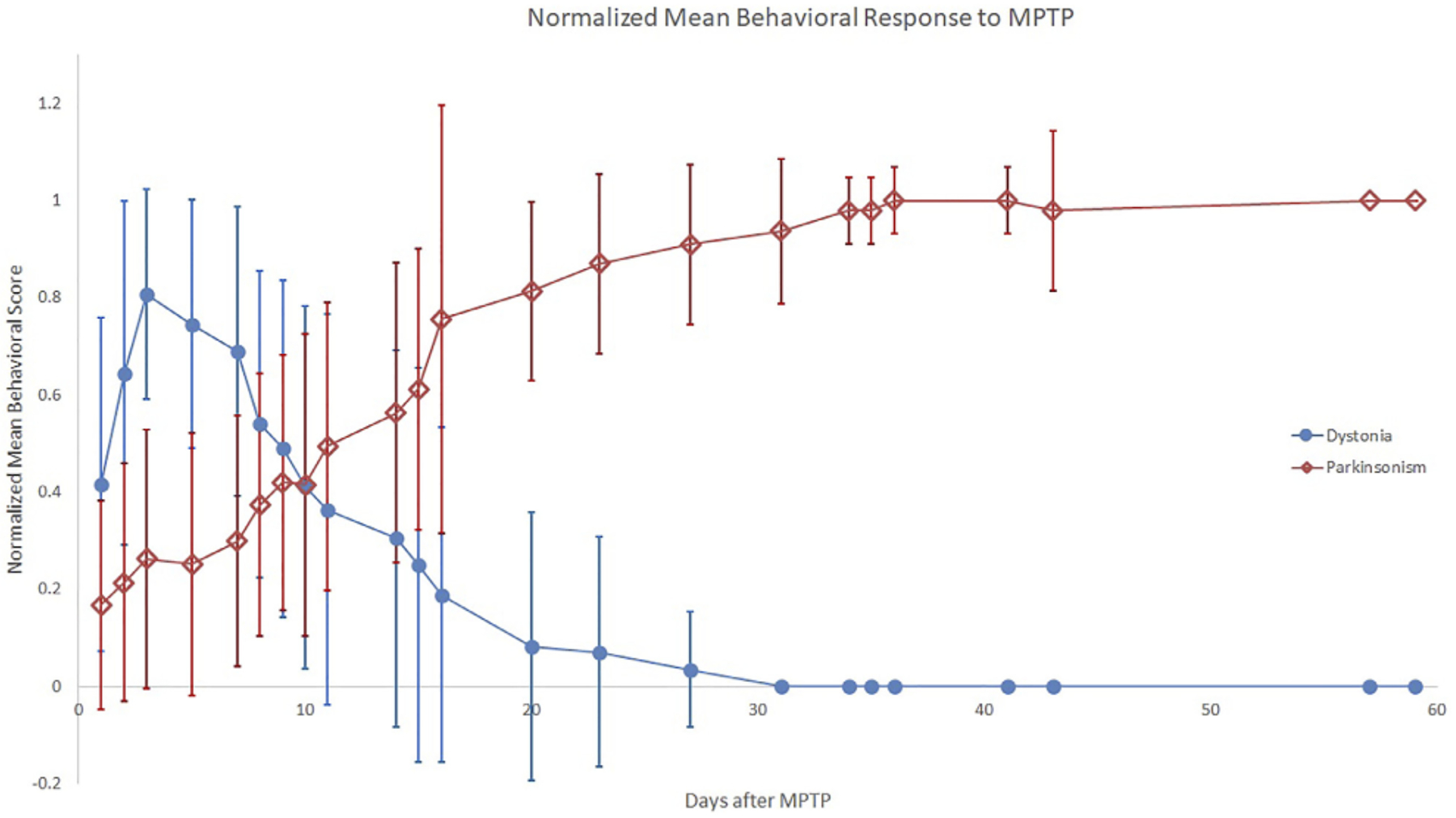
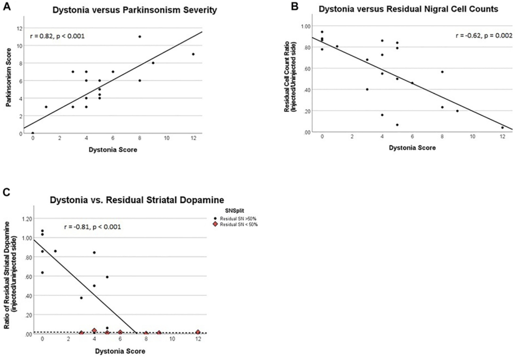
Unilateral internal carotid artery 1-methyl-4-phenyl-1,2,3,6-tetrahydropyridine (MPTP) infusion in non-human primates produces transient contralateral hemi-dystonia followed by stable contralateral hemi-parkinsonism; the relationship between dystonia and parkinsonism remains unclear. We hypothesized that transient dystonia severity following MPTP correlates with parkinsonism severity. In male Macaca nemestrina (n = 3) and M. fascicularis (n = 17) we administered unilateral intra-carotid MPTP, then correlated validated blinded ratings of transient peak dystonia and delayed parkinsonism. We also correlated dystonia severity with post-mortem measures of residual striatal dopamine and nigral neuron counts obtained a mean 53 ± 15 days following MPTP, after resolution of dystonia but during stable parkinsonism. Median latency to dystonia onset was 1 day, and peak severity 2.5 days after MPTP; total dystonia duration was 13.5 days. Parkinsonism peaked a median of 19.5 days after MPTP, remaining nearly constant thereafter. Peak dystonia severity highly correlated with parkinsonism severity (r[18] = 0.82, p < 0.001). Residual cell counts in lesioned nigra correlated linearly with peak dystonia scores (r[18] = -0.68, p=<0.001). Dystonia was not observed in monkeys without striatal dopamine depletion (n = 2); dystonia severity correlated with striatal dopamine depletion when residual nigral cell loss was less than 50% ([11] r = -0.83, p < 0.001) but spanned a broad range with near complete striatal dopamine depletion, when nigral cell loss was greater than 50%. Our data indicate that residual striatal dopamine may not reflect dystonia severity. We speculate on mechanisms of transient dystonia followed by parkinsonism that may be studied using this particular NHP MPTP model to better understand relationships of transient dystonia to nigrostriatal injury and parkinsonism.


 分享
分享

 求助内容:
求助内容: 应助结果提醒方式:
 扫码关注我们
