{"title":"Anatomical identification of a corticocortical top-down recipient inhibitory circuitry by enhancer-restricted transsynaptic tracing.","authors":"Yusuke Atsumi, Yasuhiro Oisi, Maya Odagawa, Chie Matsubara, Yoshihito Saito, Hiroyuki Uwamori, Kenta Kobayashi, Shigeki Kato, Kazuto Kobayashi, Masanori Murayama","doi":"10.3389/fncir.2023.1245097","DOIUrl":null,"url":null,"abstract":"<p><p>Despite the importance of postsynaptic inhibitory circuitry targeted by mid/long-range projections (e.g., top-down projections) in cognitive functions, its anatomical properties, such as laminar profile and neuron type, are poorly understood owing to the lack of efficient tracing methods. To this end, we developed a method that combines conventional adeno-associated virus (AAV)-mediated transsynaptic tracing with a distal-less homeobox (Dlx) enhancer-restricted expression system to label postsynaptic inhibitory neurons. We called this method \"Dlx enhancer-restricted Interneuron-SpECific transsynaptic Tracing\" (DISECT). We applied DISECT to a top-down corticocortical circuit from the secondary motor cortex (M2) to the primary somatosensory cortex (S1) in wild-type mice. First, we injected AAV1-Cre into the M2, which enabled Cre recombinase expression in M2-input recipient S1 neurons. Second, we injected AAV1-hDlx-flex-green fluorescent protein (GFP) into the S1 to transduce GFP into the postsynaptic inhibitory neurons in a Cre-dependent manner. We succeeded in exclusively labeling the recipient inhibitory neurons in the S1. Laminar profile analysis of the neurons labeled via DISECT indicated that the M2-input recipient inhibitory neurons were distributed in the superficial and deep layers of the S1. This laminar distribution was aligned with the laminar density of axons projecting from the M2. We further classified the labeled neuron types using immunohistochemistry and <i>in situ</i> hybridization. This <i>post hoc</i> classification revealed that the dominant top-down M2-input recipient neuron types were somatostatin-expressing neurons in the superficial layers and parvalbumin-expressing neurons in the deep layers. These results demonstrate that DISECT enables the investigation of multiple anatomical properties of the postsynaptic inhibitory circuitry.</p>","PeriodicalId":12498,"journal":{"name":"Frontiers in Neural Circuits","volume":"17 ","pages":"1245097"},"PeriodicalIF":3.0000,"publicationDate":"2023-08-30","publicationTypes":"Journal Article","fieldsOfStudy":null,"isOpenAccess":false,"openAccessPdf":"https://www.ncbi.nlm.nih.gov/pmc/articles/PMC10502327/pdf/","citationCount":"0","resultStr":null,"platform":"Semanticscholar","paperid":null,"PeriodicalName":"Frontiers in Neural Circuits","FirstCategoryId":"3","ListUrlMain":"https://doi.org/10.3389/fncir.2023.1245097","RegionNum":3,"RegionCategory":"医学","ArticlePicture":[],"TitleCN":null,"AbstractTextCN":null,"PMCID":null,"EPubDate":"2023/1/1 0:00:00","PubModel":"eCollection","JCR":"Q2","JCRName":"NEUROSCIENCES","Score":null,"Total":0}
引用次数: 0
Abstract
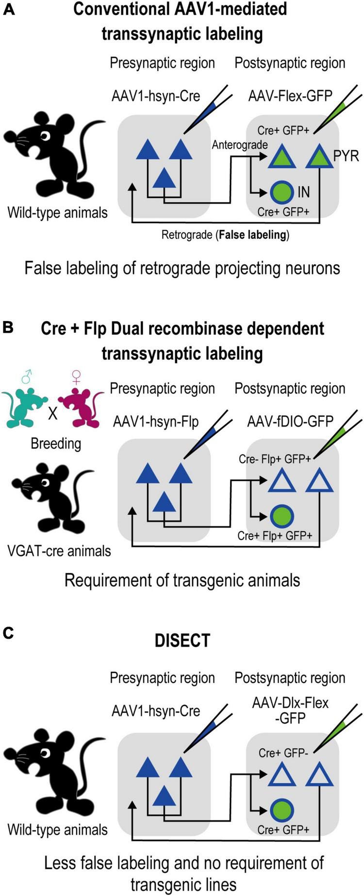
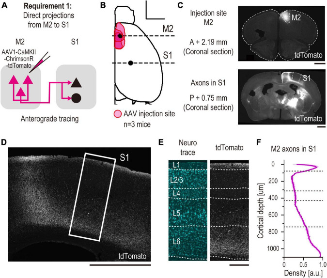
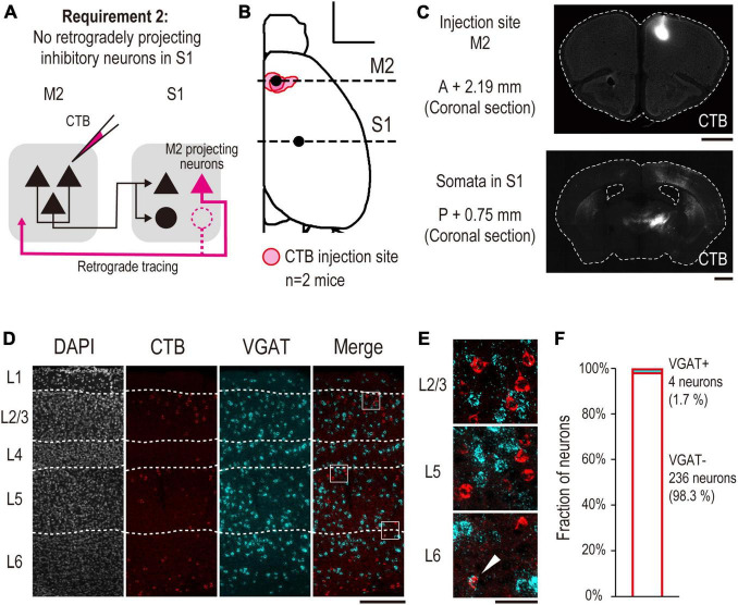
Despite the importance of postsynaptic inhibitory circuitry targeted by mid/long-range projections (e.g., top-down projections) in cognitive functions, its anatomical properties, such as laminar profile and neuron type, are poorly understood owing to the lack of efficient tracing methods. To this end, we developed a method that combines conventional adeno-associated virus (AAV)-mediated transsynaptic tracing with a distal-less homeobox (Dlx) enhancer-restricted expression system to label postsynaptic inhibitory neurons. We called this method "Dlx enhancer-restricted Interneuron-SpECific transsynaptic Tracing" (DISECT). We applied DISECT to a top-down corticocortical circuit from the secondary motor cortex (M2) to the primary somatosensory cortex (S1) in wild-type mice. First, we injected AAV1-Cre into the M2, which enabled Cre recombinase expression in M2-input recipient S1 neurons. Second, we injected AAV1-hDlx-flex-green fluorescent protein (GFP) into the S1 to transduce GFP into the postsynaptic inhibitory neurons in a Cre-dependent manner. We succeeded in exclusively labeling the recipient inhibitory neurons in the S1. Laminar profile analysis of the neurons labeled via DISECT indicated that the M2-input recipient inhibitory neurons were distributed in the superficial and deep layers of the S1. This laminar distribution was aligned with the laminar density of axons projecting from the M2. We further classified the labeled neuron types using immunohistochemistry and in situ hybridization. This post hoc classification revealed that the dominant top-down M2-input recipient neuron types were somatostatin-expressing neurons in the superficial layers and parvalbumin-expressing neurons in the deep layers. These results demonstrate that DISECT enables the investigation of multiple anatomical properties of the postsynaptic inhibitory circuitry.
期刊介绍:
Frontiers in Neural Circuits publishes rigorously peer-reviewed research on the emergent properties of neural circuits - the elementary modules of the brain. Specialty Chief Editors Takao K. Hensch and Edward Ruthazer at Harvard University and McGill University respectively, are supported by an outstanding Editorial Board of international experts. This multidisciplinary open-access journal is at the forefront of disseminating and communicating scientific knowledge and impactful discoveries to researchers, academics and the public worldwide.
Frontiers in Neural Circuits launched in 2011 with great success and remains a "central watering hole" for research in neural circuits, serving the community worldwide to share data, ideas and inspiration. Articles revealing the anatomy, physiology, development or function of any neural circuitry in any species (from sponges to humans) are welcome. Our common thread seeks the computational strategies used by different circuits to link their structure with function (perceptual, motor, or internal), the general rules by which they operate, and how their particular designs lead to the emergence of complex properties and behaviors. Submissions focused on synaptic, cellular and connectivity principles in neural microcircuits using multidisciplinary approaches, especially newer molecular, developmental and genetic tools, are encouraged. Studies with an evolutionary perspective to better understand how circuit design and capabilities evolved to produce progressively more complex properties and behaviors are especially welcome. The journal is further interested in research revealing how plasticity shapes the structural and functional architecture of neural circuits.


 分享
分享
 求助内容:
求助内容: 应助结果提醒方式:
 扫码关注我们
