Devkumar Mustafi, Abby Leinroth, Xiaobing Fan, Erica Markiewicz, Marta Zamora, Jeffrey Mueller, Suzanne D Conzen, Gregory S Karczmar
{"title":"Magnetic Resonance Angiography Shows Increased Arterial Blood Supply Associated with Murine Mammary Cancer.","authors":"Devkumar Mustafi, Abby Leinroth, Xiaobing Fan, Erica Markiewicz, Marta Zamora, Jeffrey Mueller, Suzanne D Conzen, Gregory S Karczmar","doi":"10.1155/2019/5987425","DOIUrl":null,"url":null,"abstract":"Breast cancer is a major cause of morbidity and mortality in Western women. Tumor neoangiogenesis, the formation of new blood vessels from pre-existing ones, may be used as a prognostic marker for cancer progression. Clinical practice uses dynamic contrast enhanced magnetic resonance imaging (DCE-MRI) to detect cancers based on increased blood flow and capillary permeability. However, DCE-MRI requires repeated injections of contrast media. Therefore we explored the use of noninvasive time-of-flight (TOF) MR angiography for serial studies of mouse mammary glands to measure the number and size of arteries feeding mammary glands with and without cancer. Virgin female C3(1) SV40 TAg mice (n=9), aged 18-20 weeks, were imaged on a 9.4 Tesla small animal scanner. Multislice T2-weighted (T2W) images and TOF-MRI angiograms were acquired over inguinal mouse mammary glands. The data were analyzed to determine tumor burden in each mammary gland and the volume of arteries feeding each mammary gland. After in vivo MRI, inguinal mammary glands were excised and fixed in formalin for histology. TOF angiography detected arteries with a diameter as small as 0.1 mm feeding the mammary glands. A significant correlation (r=0.79; p< 0.0001) was found between tumor volume and the arterial blood volume measured in mammary glands. Mammary arterial blood volumes ranging from 0.08 mm3 to 3.81 mm3 were measured. Tumors and blood vessels found on in vivo T2W and TOF images, respectively, were confirmed with ex vivo histological images. These results demonstrate increased recruitment of arteries to mammary glands with cancer, likely associated with neoangiogenesis. Neoangiogenesis may be detected by TOF angiography without injection of contrast agents. This would be very useful in mouse models where repeat placement of I.V. lines is challenging. In addition, analogous methods could be tested in humans to evaluate the vasculature of suspicious lesions without using contrast agents.","PeriodicalId":47063,"journal":{"name":"International Journal of Biomedical Imaging","volume":"2019 ","pages":"5987425"},"PeriodicalIF":1.3000,"publicationDate":"2019-01-01","publicationTypes":"Journal Article","fieldsOfStudy":null,"isOpenAccess":false,"openAccessPdf":"https://sci-hub-pdf.com/10.1155/2019/5987425","citationCount":"4","resultStr":null,"platform":"Semanticscholar","paperid":null,"PeriodicalName":"International Journal of Biomedical Imaging","FirstCategoryId":"1085","ListUrlMain":"https://doi.org/10.1155/2019/5987425","RegionNum":0,"RegionCategory":null,"ArticlePicture":[],"TitleCN":null,"AbstractTextCN":null,"PMCID":null,"EPubDate":"","PubModel":"","JCR":"Q2","JCRName":"ENGINEERING, BIOMEDICAL","Score":null,"Total":0}
引用次数: 4
Abstract
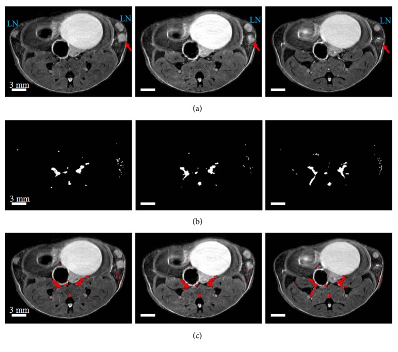
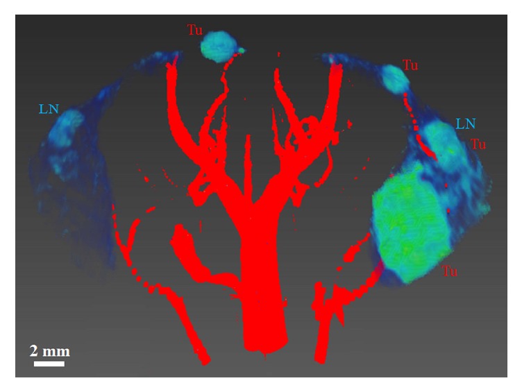
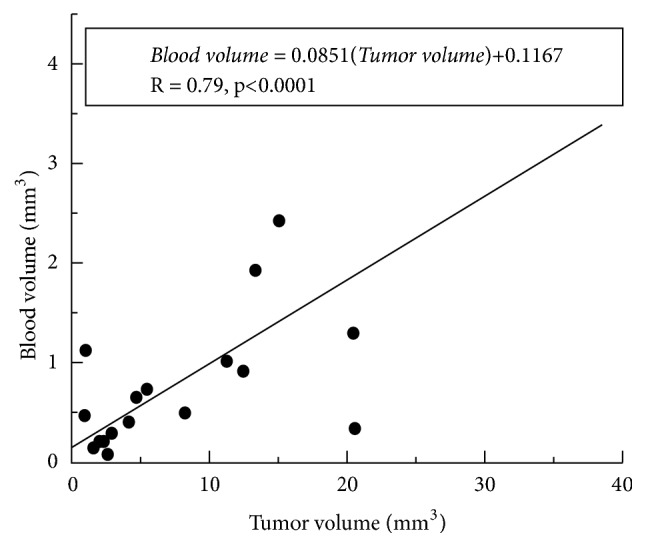
Breast cancer is a major cause of morbidity and mortality in Western women. Tumor neoangiogenesis, the formation of new blood vessels from pre-existing ones, may be used as a prognostic marker for cancer progression. Clinical practice uses dynamic contrast enhanced magnetic resonance imaging (DCE-MRI) to detect cancers based on increased blood flow and capillary permeability. However, DCE-MRI requires repeated injections of contrast media. Therefore we explored the use of noninvasive time-of-flight (TOF) MR angiography for serial studies of mouse mammary glands to measure the number and size of arteries feeding mammary glands with and without cancer. Virgin female C3(1) SV40 TAg mice (n=9), aged 18-20 weeks, were imaged on a 9.4 Tesla small animal scanner. Multislice T2-weighted (T2W) images and TOF-MRI angiograms were acquired over inguinal mouse mammary glands. The data were analyzed to determine tumor burden in each mammary gland and the volume of arteries feeding each mammary gland. After in vivo MRI, inguinal mammary glands were excised and fixed in formalin for histology. TOF angiography detected arteries with a diameter as small as 0.1 mm feeding the mammary glands. A significant correlation (r=0.79; p< 0.0001) was found between tumor volume and the arterial blood volume measured in mammary glands. Mammary arterial blood volumes ranging from 0.08 mm3 to 3.81 mm3 were measured. Tumors and blood vessels found on in vivo T2W and TOF images, respectively, were confirmed with ex vivo histological images. These results demonstrate increased recruitment of arteries to mammary glands with cancer, likely associated with neoangiogenesis. Neoangiogenesis may be detected by TOF angiography without injection of contrast agents. This would be very useful in mouse models where repeat placement of I.V. lines is challenging. In addition, analogous methods could be tested in humans to evaluate the vasculature of suspicious lesions without using contrast agents.
期刊介绍:
The International Journal of Biomedical Imaging is managed by a board of editors comprising internationally renowned active researchers. The journal is freely accessible online and also offered for purchase in print format. It employs a web-based review system to ensure swift turnaround times while maintaining high standards. In addition to regular issues, special issues are organized by guest editors. The subject areas covered include (but are not limited to):
Digital radiography and tomosynthesis
X-ray computed tomography (CT)
Magnetic resonance imaging (MRI)
Single photon emission computed tomography (SPECT)
Positron emission tomography (PET)
Ultrasound imaging
Diffuse optical tomography, coherence, fluorescence, bioluminescence tomography, impedance tomography
Neutron imaging for biomedical applications
Magnetic and optical spectroscopy, and optical biopsy
Optical, electron, scanning tunneling/atomic force microscopy
Small animal imaging
Functional, cellular, and molecular imaging
Imaging assays for screening and molecular analysis
Microarray image analysis and bioinformatics
Emerging biomedical imaging techniques
Imaging modality fusion
Biomedical imaging instrumentation
Biomedical image processing, pattern recognition, and analysis
Biomedical image visualization, compression, transmission, and storage
Imaging and modeling related to systems biology and systems biomedicine
Applied mathematics, applied physics, and chemistry related to biomedical imaging
Grid-enabling technology for biomedical imaging and informatics


 分享
分享
 求助内容:
求助内容: 应助结果提醒方式:
 扫码关注我们
