{"title":"Crystal structure of Sphingobacterium multivorum serine palmitoyltransferase complexed with tris(hydroxymethyl)aminomethane","authors":"Hiroko Ikushiro, Aya Takahashi, Taiki Murakami, Asuka Katayama, Taiki Sawai, Haruna Goto, Ikuko Miyahara, Nobuo Kamiya, Takato Yano","doi":"10.1107/S2053230X22010937","DOIUrl":null,"url":null,"abstract":"<p>Serine palmitoyltransferase (SPT) catalyses the first reaction in sphingolipid biosynthesis: the decarboxylative condensation of <span>l</span>-serine (<span>l</span>-Ser) and palmitoyl-CoA to form 3-ketodihydrosphingosine. SPT from <i>Sphingobacterium multivorum</i> has been isolated and its crystal structure in complex with <span>l</span>-Ser has been determined at 2.3 Å resolution (PDB entry 3a2b). However, the quality of the crystal was not good enough to judge the conformation of the cofactor molecule and the orientations of the side chains of the amino-acid residues in the enzyme active site. The crystal quality was improved by revision of the purification procedure and by optimization of both the crystallization procedure and the post-crystallization treatment conditions. Here, the crystal structure of SPT complexed with tris(hydroxymethyl)aminomethane (Tris), a buffer component, was determined at 1.65 Å resolution. The protein crystallized at 20°C and diffraction data were collected from the crystals to a resolution of 1.65 Å. The crystal belonged to the tetragonal space group <i>P</i>4<sub>1</sub>2<sub>1</sub>2, with unit-cell parameters <i>a</i> = <i>b</i> = 61.32, <i>c</i> = 208.57 Å. Analysis of the crystal structure revealed C4—C5—C5A—O4P (77°) and C5—C5A—O4P—P (–143°) torsion angles in the phosphate-group moiety of the cofactor pyridoxal 5′-phosphate (PLP) that are more reasonable than those observed in the previously reported crystal structure (14° and 151°, respectively). Furthermore, the clear electron density showing a Schiff-base linkage between PLP and the bulky artificial ligand Tris indicated exceptional flexibility of the active-site cavity of this enzyme. These findings open up the possibility for further study of the detailed mechanisms of substrate recognition and catalysis by this enzyme.</p>","PeriodicalId":7029,"journal":{"name":"Acta crystallographica. Section F, Structural biology communications","volume":"78 12","pages":"408-415"},"PeriodicalIF":1.1000,"publicationDate":"2022-11-28","publicationTypes":"Journal Article","fieldsOfStudy":null,"isOpenAccess":false,"openAccessPdf":"","citationCount":"1","resultStr":null,"platform":"Semanticscholar","paperid":null,"PeriodicalName":"Acta crystallographica. Section F, Structural biology communications","FirstCategoryId":"99","ListUrlMain":"https://onlinelibrary.wiley.com/doi/10.1107/S2053230X22010937","RegionNum":4,"RegionCategory":"生物学","ArticlePicture":[],"TitleCN":null,"AbstractTextCN":null,"PMCID":null,"EPubDate":"","PubModel":"","JCR":"Q4","JCRName":"BIOCHEMICAL RESEARCH METHODS","Score":null,"Total":0}
引用次数: 1
Abstract
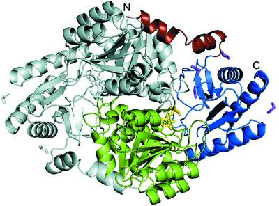
Serine palmitoyltransferase (SPT) catalyses the first reaction in sphingolipid biosynthesis: the decarboxylative condensation of l-serine (l-Ser) and palmitoyl-CoA to form 3-ketodihydrosphingosine. SPT from Sphingobacterium multivorum has been isolated and its crystal structure in complex with l-Ser has been determined at 2.3 Å resolution (PDB entry 3a2b). However, the quality of the crystal was not good enough to judge the conformation of the cofactor molecule and the orientations of the side chains of the amino-acid residues in the enzyme active site. The crystal quality was improved by revision of the purification procedure and by optimization of both the crystallization procedure and the post-crystallization treatment conditions. Here, the crystal structure of SPT complexed with tris(hydroxymethyl)aminomethane (Tris), a buffer component, was determined at 1.65 Å resolution. The protein crystallized at 20°C and diffraction data were collected from the crystals to a resolution of 1.65 Å. The crystal belonged to the tetragonal space group P41212, with unit-cell parameters a = b = 61.32, c = 208.57 Å. Analysis of the crystal structure revealed C4—C5—C5A—O4P (77°) and C5—C5A—O4P—P (–143°) torsion angles in the phosphate-group moiety of the cofactor pyridoxal 5′-phosphate (PLP) that are more reasonable than those observed in the previously reported crystal structure (14° and 151°, respectively). Furthermore, the clear electron density showing a Schiff-base linkage between PLP and the bulky artificial ligand Tris indicated exceptional flexibility of the active-site cavity of this enzyme. These findings open up the possibility for further study of the detailed mechanisms of substrate recognition and catalysis by this enzyme.
期刊介绍:
Acta Crystallographica Section F is a rapid structural biology communications journal.
Articles on any aspect of structural biology, including structures determined using high-throughput methods or from iterative studies such as those used in the pharmaceutical industry, are welcomed by the journal.
The journal offers the option of open access, and all communications benefit from unlimited free use of colour illustrations and no page charges. Authors are encouraged to submit multimedia content for publication with their articles.
Acta Cryst. F has a dedicated online tool called publBio that is designed to make the preparation and submission of articles easier for authors.


 分享
分享
 求助内容:
求助内容: 应助结果提醒方式:
 扫码关注我们
