Evaluation of the contour of edentulous jaw sections in the transversal plane and the buccolingual vertical-level disparity in CBCT and panoramic radiography images: a retrospective comparative study.
Ali Reza Ketabi, Andree Piwowarczyk, Matthias Christian Schulz, Hans-Christoph Lauer, Stefan Hassfeld
{"title":"Evaluation of the contour of edentulous jaw sections in the transversal plane and the buccolingual vertical-level disparity in CBCT and panoramic radiography images: a retrospective comparative study.","authors":"Ali Reza Ketabi, Andree Piwowarczyk, Matthias Christian Schulz, Hans-Christoph Lauer, Stefan Hassfeld","doi":"10.1186/s40729-022-00466-8","DOIUrl":null,"url":null,"abstract":"<p><strong>Purpose: </strong>This study investigates whether edentulous jaw sections in the planned implant position exhibit jaw contours funnel-shaped or exhibit pronounced retraction of the jaw (unusual jaw contours) in the transversal plane of the three-dimensional (3D) images, not visible in two-dimensional (2D) images.</p><p><strong>Methods: </strong>A total of 335 patients with an edentulous section of the jaw that required dental implants were selected. Anonymised radiologic patients' data were collected, comprising cone-beam computed tomography (CBCT) images of the edentulous jaw sections. In the first stage, unusual jaw contours were examined, including funnel-shaped or pronounced retraction of the jaw and hypodense regions with an undercut and/or bone deficit. In the second stage, the variation in the height of the alveolar ridge between the lingual and buccal contour in the edentulous jaw sections was assessed.</p><p><strong>Results: </strong>The CBCT images of an unusual jaw contour were observed in 8 cases (2.4%) in the maxilla on the left and 10 cases (3%) in the maxilla on the right. In the mandible, a jaw contour deviates in 39 cases (12.1%) on the left side and 39 cases (12.1%) on the right side. A height difference was detected in the upper jaw in 307 cases and the lower jaw in 265 cases. The discrepancy was 2.09 mm (± 2.25 mm) in the maxilla and 3.97 mm (± 3.45 mm) in the mandible.</p><p><strong>Conclusions: </strong>The CBCT scan provides useful information to avoid complications in the preoperative planning phase and surgical planning in implant dentistry.</p>","PeriodicalId":14076,"journal":{"name":"International Journal of Implant Dentistry","volume":"9 1","pages":"1"},"PeriodicalIF":4.0000,"publicationDate":"2023-01-03","publicationTypes":"Journal Article","fieldsOfStudy":null,"isOpenAccess":false,"openAccessPdf":"https://www.ncbi.nlm.nih.gov/pmc/articles/PMC9810779/pdf/","citationCount":"0","resultStr":null,"platform":"Semanticscholar","paperid":null,"PeriodicalName":"International Journal of Implant Dentistry","FirstCategoryId":"3","ListUrlMain":"https://doi.org/10.1186/s40729-022-00466-8","RegionNum":3,"RegionCategory":"医学","ArticlePicture":[],"TitleCN":null,"AbstractTextCN":null,"PMCID":null,"EPubDate":"","PubModel":"","JCR":"Q1","JCRName":"DENTISTRY, ORAL SURGERY & MEDICINE","Score":null,"Total":0}
引用次数: 0
Abstract
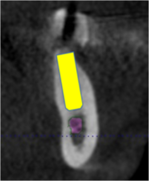
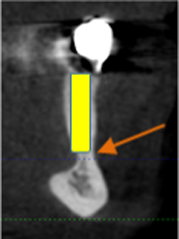
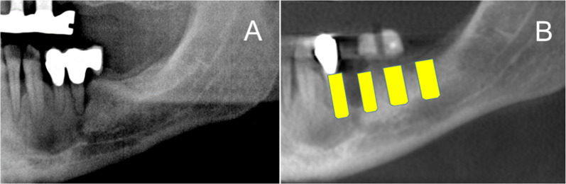
Purpose: This study investigates whether edentulous jaw sections in the planned implant position exhibit jaw contours funnel-shaped or exhibit pronounced retraction of the jaw (unusual jaw contours) in the transversal plane of the three-dimensional (3D) images, not visible in two-dimensional (2D) images.
Methods: A total of 335 patients with an edentulous section of the jaw that required dental implants were selected. Anonymised radiologic patients' data were collected, comprising cone-beam computed tomography (CBCT) images of the edentulous jaw sections. In the first stage, unusual jaw contours were examined, including funnel-shaped or pronounced retraction of the jaw and hypodense regions with an undercut and/or bone deficit. In the second stage, the variation in the height of the alveolar ridge between the lingual and buccal contour in the edentulous jaw sections was assessed.
Results: The CBCT images of an unusual jaw contour were observed in 8 cases (2.4%) in the maxilla on the left and 10 cases (3%) in the maxilla on the right. In the mandible, a jaw contour deviates in 39 cases (12.1%) on the left side and 39 cases (12.1%) on the right side. A height difference was detected in the upper jaw in 307 cases and the lower jaw in 265 cases. The discrepancy was 2.09 mm (± 2.25 mm) in the maxilla and 3.97 mm (± 3.45 mm) in the mandible.
Conclusions: The CBCT scan provides useful information to avoid complications in the preoperative planning phase and surgical planning in implant dentistry.
期刊介绍:
The International Journal of Implant Dentistry is a peer-reviewed open access journal published under the SpringerOpen brand. The journal is dedicated to promoting the exchange and discussion of all research areas relevant to implant dentistry in the form of systematic literature or invited reviews, prospective and retrospective clinical studies, clinical case reports, basic laboratory and animal research, and articles on material research and engineering.


 分享
分享
 求助内容:
求助内容: 应助结果提醒方式:
 扫码关注我们
