{"title":"Regulation of Collagen I and Collagen III in Tissue Injury and Regeneration.","authors":"Drishtant Singh, Vikrant Rai, Devendra K Agrawal","doi":"10.26502/fccm.92920302","DOIUrl":null,"url":null,"abstract":"<p><p>The structure of connective tissues including cartilage, tendons, and ligaments as well as many organs, like the skin, heart, liver, kidney, lungs, blood vessels, and bones, depend on collagen. The bulk of the network of structural proteins that make up the extracellular matrix of the heart is composed of collagen type I and type III, which provide structural support for the muscle cells and are crucial for cardiac function. The prognosis and progression of a disease or diseased state may be significantly impacted by the upregulation or downregulation of the collagen types, particularly Col I and Col III. For example, increasing Col I protein levels may impose increasing myocardial stiffness, impairing the diastolic and systolic function of the myocardium. Collagen I is a stiff fibrillar protein that gives tensile strength, whereas Col III produces an elastic network that stores kinetic energy as an elastic rebound. These two collagen proteins have distinct physical properties in nature. Therefore, the control of Col I and Col III as well as the potential relevance of the Col I/Col III ratio in many biological processes serve as the foundation for this comprehensive review article.</p>","PeriodicalId":72523,"journal":{"name":"Cardiology and cardiovascular medicine","volume":"7 1","pages":"5-16"},"PeriodicalIF":0.0000,"publicationDate":"2023-01-01","publicationTypes":"Journal Article","fieldsOfStudy":null,"isOpenAccess":false,"openAccessPdf":"https://www.ncbi.nlm.nih.gov/pmc/articles/PMC9912297/pdf/","citationCount":"14","resultStr":null,"platform":"Semanticscholar","paperid":null,"PeriodicalName":"Cardiology and cardiovascular medicine","FirstCategoryId":"1085","ListUrlMain":"https://doi.org/10.26502/fccm.92920302","RegionNum":0,"RegionCategory":null,"ArticlePicture":[],"TitleCN":null,"AbstractTextCN":null,"PMCID":null,"EPubDate":"","PubModel":"","JCR":"","JCRName":"","Score":null,"Total":0}
引用次数: 14
Abstract
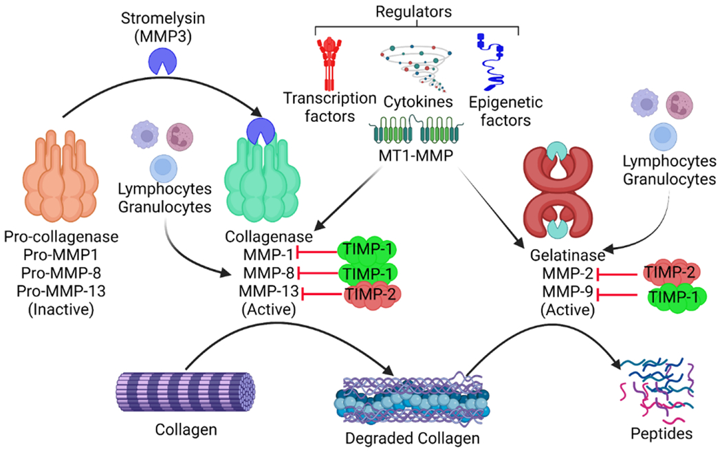
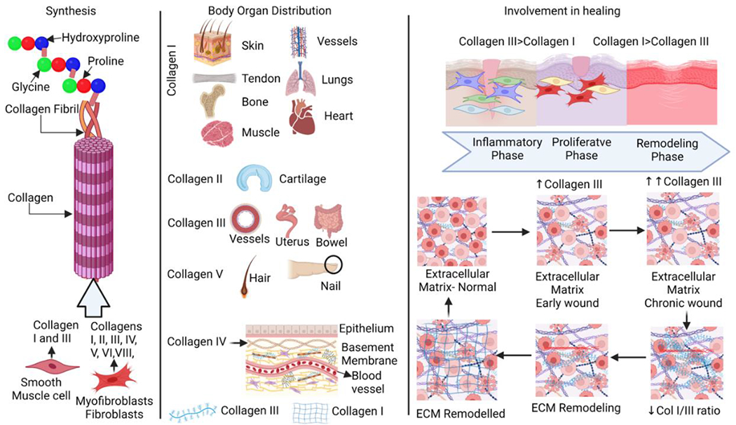
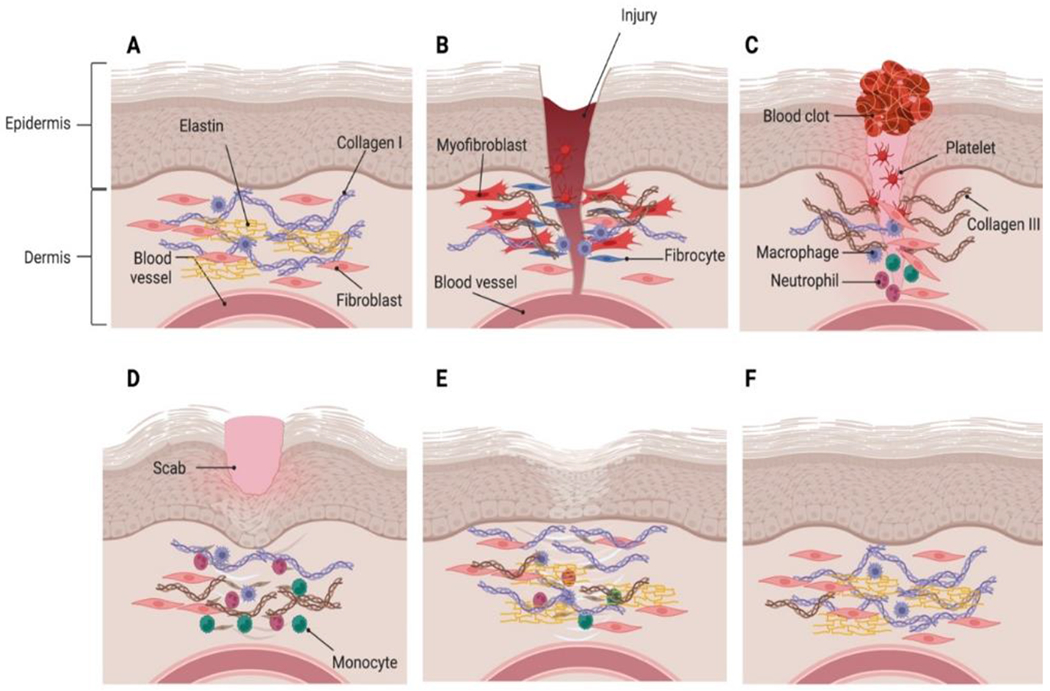
The structure of connective tissues including cartilage, tendons, and ligaments as well as many organs, like the skin, heart, liver, kidney, lungs, blood vessels, and bones, depend on collagen. The bulk of the network of structural proteins that make up the extracellular matrix of the heart is composed of collagen type I and type III, which provide structural support for the muscle cells and are crucial for cardiac function. The prognosis and progression of a disease or diseased state may be significantly impacted by the upregulation or downregulation of the collagen types, particularly Col I and Col III. For example, increasing Col I protein levels may impose increasing myocardial stiffness, impairing the diastolic and systolic function of the myocardium. Collagen I is a stiff fibrillar protein that gives tensile strength, whereas Col III produces an elastic network that stores kinetic energy as an elastic rebound. These two collagen proteins have distinct physical properties in nature. Therefore, the control of Col I and Col III as well as the potential relevance of the Col I/Col III ratio in many biological processes serve as the foundation for this comprehensive review article.


 分享
分享

 求助内容:
求助内容: 应助结果提醒方式:
 扫码关注我们
