Irregular Baseline Brain Activity in Coronary Artery Disease Patients with Cognitive Impairment: A Resting-state Functional Magnetic Resonance Imaging Study.
{"title":"Irregular Baseline Brain Activity in Coronary Artery Disease Patients with Cognitive Impairment: A Resting-state Functional Magnetic Resonance Imaging Study.","authors":"Jingchen Zhang, Jueyue Yan, Jianhua Niu, Zhipeng Xu, Xing Fang, Jingyu You, Tong Li","doi":"10.2174/1567202619666220516124552","DOIUrl":null,"url":null,"abstract":"Objective Cognitive impairment has been suggested to be associated with coronary artery disease [CAD]; however, the underlying mechanism is not fully understood. Our current study aimed to explore the brain activity in CAD patients compared to healthy controls [HCs]. Methods Twenty-two CAD patients and 23 HCs were enrolled in our study. A low-frequency oscillation at the voxel level in all participants based on the amplitude of low-frequency fluctuations [ALFF] was measured using resting-state functional magnetic resonance imaging. All participants underwent neuropsychological examinations [Mini-Mental State Examination, MMSE and Montreal Cognitive Assessment, MoCA] and visual acuity examination. Results CAD patients showed significantly lower ALFF values [P < 0.05] in the right precuneus gyrus [Precuneus_R], left supramarginal gyrus [Supramarginal_L], left angular gyrus [Angular_L], and left middle cingulum gyrus [Cingulum_Mid_L] than healthy controls. Lower MoCA scores in CAD patients significantly correlated with lower Supramarginal_L [P = 0.001] and Cingulate_Mid_L [P = 0.004] ALFF values. Reduced visual acuity significantly correlated with lower Precuneus_R [P = 0.019] and Cingulate_Mid_L [P = 0.011] ALFF values in CAD patients. Conclusion These findings may provide further insight into the underlying neuropathophysiology of CAD with cognitive impairment.","PeriodicalId":10879,"journal":{"name":"Current neurovascular research","volume":"19 2","pages":"131-136"},"PeriodicalIF":1.7000,"publicationDate":"2022-01-01","publicationTypes":"Journal Article","fieldsOfStudy":null,"isOpenAccess":false,"openAccessPdf":"https://www.ncbi.nlm.nih.gov/pmc/articles/PMC9933043/pdf/","citationCount":"3","resultStr":null,"platform":"Semanticscholar","paperid":null,"PeriodicalName":"Current neurovascular research","FirstCategoryId":"3","ListUrlMain":"https://doi.org/10.2174/1567202619666220516124552","RegionNum":4,"RegionCategory":"医学","ArticlePicture":[],"TitleCN":null,"AbstractTextCN":null,"PMCID":null,"EPubDate":"","PubModel":"","JCR":"Q3","JCRName":"CLINICAL NEUROLOGY","Score":null,"Total":0}
引用次数: 3
Abstract
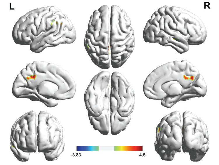
Objective Cognitive impairment has been suggested to be associated with coronary artery disease [CAD]; however, the underlying mechanism is not fully understood. Our current study aimed to explore the brain activity in CAD patients compared to healthy controls [HCs]. Methods Twenty-two CAD patients and 23 HCs were enrolled in our study. A low-frequency oscillation at the voxel level in all participants based on the amplitude of low-frequency fluctuations [ALFF] was measured using resting-state functional magnetic resonance imaging. All participants underwent neuropsychological examinations [Mini-Mental State Examination, MMSE and Montreal Cognitive Assessment, MoCA] and visual acuity examination. Results CAD patients showed significantly lower ALFF values [P < 0.05] in the right precuneus gyrus [Precuneus_R], left supramarginal gyrus [Supramarginal_L], left angular gyrus [Angular_L], and left middle cingulum gyrus [Cingulum_Mid_L] than healthy controls. Lower MoCA scores in CAD patients significantly correlated with lower Supramarginal_L [P = 0.001] and Cingulate_Mid_L [P = 0.004] ALFF values. Reduced visual acuity significantly correlated with lower Precuneus_R [P = 0.019] and Cingulate_Mid_L [P = 0.011] ALFF values in CAD patients. Conclusion These findings may provide further insight into the underlying neuropathophysiology of CAD with cognitive impairment.
期刊介绍:
Current Neurovascular Research provides a cross platform for the publication of scientifically rigorous research that addresses disease mechanisms of both neuronal and vascular origins in neuroscience. The journal serves as an international forum publishing novel and original work as well as timely neuroscience research articles, full-length/mini reviews in the disciplines of cell developmental disorders, plasticity, and degeneration that bridges the gap between basic science research and clinical discovery. Current Neurovascular Research emphasizes the elucidation of disease mechanisms, both cellular and molecular, which can impact the development of unique therapeutic strategies for neuronal and vascular disorders.


 分享
分享
 求助内容:
求助内容: 应助结果提醒方式:
 扫码关注我们
